Chapter 42 Rheumatoid Arthritis
Conservative Surgical Procedures Open/Arthroscopic Synovectomy
Introduction
Involvement of the elbow is rarely the initial presenting sign of rheumatoid arthritis, nor does it usually present in isolation. The clinical signs in elbow pathology generally occur with more extensive disease, affecting up to 60% of the rheumatoid population.1 Patients often present late, and the options for treatment can be dramatically reduced due to age, disease progression and post-management rehabilitation requirements.
Background/aetiology
Recent advances in medical management (especially the anti-tumour necrosis factor (TNF) α drugs including etanercept (Enbrel), infliximab (Remicade) and adalimumab (Humira)) have significantly slowed the rate of disease progression and therefore dramatically reduced the number of patients presenting with severe rheumatoid joint deformity and destruction.2 Consequently early detection of the disease and medical intervention where appropriate have become a priority.
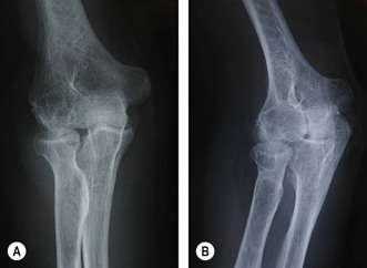
The presentation of rheumatoid arthritis is extremely variable, with systemic symptoms sometimes complicating the picture. When joint symptoms predominate it is often a symmetrical polyarthritis. Certainly, the hands and wrists are the most frequently affected joints, with the elbow being involved in 20–60% of cases.1 Despite this variable presentation, joint involvement in rheumatoid arthritis is always characterized by inflammation and synovial hypertrophy.3 Treatment should be tailored to the pathology as it progresses through the relatively predictable radiographic Larsen stages of disease4 (Fig. 42.1). Put simply, the inflammation and synovial thickening lead to effusion, cartilage breakdown, erosions, nodules and capsulo-ligamentous thinning.
Initially alternative causes of pain, effusion and swelling in the elbow must be excluded, such as septic arthritis, posttrauma, gout and even osteoarthritis. In the earlier stages of disease the diagnosis can be difficult, especially in rheumatoid monoarthritis or pauci-articular rheumatoid arthritis, and a synovial biopsy may be desirable. Arthroscopy facilitates diagnostic biopsy, with less morbidity in experienced hands than an open procedure.5
Intraoperative findings reveal thick, villous synovium with or without rice bodies. The exudate covers the native articular cartilage from the margins, destroying the smooth edges of the cartilage and reducing the joint space.6 The often purulent joint effusion leads to friable articular cartilage in the centre of the joint, and as the elbow has a high level of constraint, this often leads to flaps of cartilage catching and/or locking the elbow as it extends. This, in addition to loose body formation, results in the common presentation of extension loss with anterior capsular tightness.
Classifications based on clinical presentation (Mayo Clinic Performance Index) and/or radiographic appearance (Mayo Clinic radiographic classification, Larsen Dale and Eek classification5) (Table 42.1) have been published by Morrey and Adams7 (as well as the elbow functional assessment (EFA) scale8 and others), but no single classification covers the variety of clinical problems produced by rheumatoid disease in the elbow.
Table 42.1 Mayo Clinic classification of the rheumatoid elbow
| Grade | Description |
|---|---|
| I | No radiographic abnormalities except periarticular osteopenia with accompanying soft tissue swelling. Mild to moderate synovitis is generally present |
| II | Mild to moderate joint space reduction with minimal or no architectural distortion. Recalcitrant synovitis that cannot be managed with non-steroidal antiinflammatory medications alone |
| III | Variable reduction in joint space with or without cyst formation. Architectural alteration, such as thinning of the olecranon, or resorption of the trochlea or capitellum. Synovitis is variable and may be quiescent |
| IV | Extensive articular damage with loss of subchondral bone and subluxation or ankylosis of the joint. Synovitis may be minimal |
From Kauffman JI, Chen AL, Stuchin S, et al. Surgical management of the rheumatoid elbow. J Am Acad Orthop Surg 2003;11:100–108; data originally from Morrey BF, Adams RA. Semiconstrained arthroplasty for the treatment of rheumatoid arthritis of the elbow. J Bone Joint Surg (Am) 1992;74:479–490.
Presentation
Articular pain, valgus deformity
The classic presentation of the severe chronic rheumatoid patient is with pain and stiffness. Pain occurs from the usual joint receptors, but also secondary to the invasive nature of the disease as there can be gross destruction of joint surfaces and of ligamentous attachments producing articular incongruity and deformity. Patients commonly present with valgus laxity due to disruption of the medial collateral ligament (MCL) as well as radial head destruction.9 Over time valgus deformity can also lead to a tardy ulnar nerve palsy.
Non-articular lesions
The proliferative synovitis may lead to synovial cyst formation. Occasionally this may also occur at the proximal radioulnar joint (PRUJ), which can result in an exertion of pressure on the posterior interosseous nerve (PIN).10 The ulnar nerve is at risk both from synovial proliferation due to its position within the cubital tunnel, and from a tardy ulnar nerve palsy from progressive valgus deformity.
Investigation
Imaging
The most reliable radiological classification for rheumatoid arthritis of the elbow is the Larson classification (Fig. 42.1). Recent advances in computed tomography (CT) have led to excellent quality three-dimensional (3D) images of the elbow to localize sites of impingement, and also to examine congruity of the joint. Magnetic resonance imaging (MRI) is, however, the investigation of choice for assessing synovitis. The presence of fluid can be distinguished from boggy synovitis, and the sites of maximum involvement can help preoperative planning and decision making.11 Ultrasound is of value when observing early synovitis and cysts, and is particularly useful when plotting the course of the radial nerve prior to surgery.12
Treatment options
Aims of these options
Surgical treatment of the elbow is used for several clinical purposes:
Table 42.2 Procedural treatment options
| Arthroscopic procedures | Arthroscopic synovial biopsy |
| Arthroscopic synovectomy | |
| Arthroscopic capsular release | |
| Arthroscopic radial head resection | |
| Open procedures | |
| Lateral or Kocher approach | Synovectomy |
| Synovectomy and capsulectomy | |
| Synovectomy/capsulectomy and radial head excision | |
| Posterior interosseous nerve release and excision of bursa | |
| Posterior approach | Olecranon nodule |
| Olecranon bursa ± spur | |
| ‘OK’ procedure | |
| Medial approach | Ulnar nerve release/transposition |
| Medial capsular release | |
| Universal incision (posterior) | |
| All of the above | Total elbow arthroplasty |
Surgical techniques of typical conservative surgical procedures
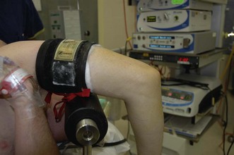
Elbow arthroscopy
Technique
If a capsulectomy is required then the safest technique is to release it off the humerus superiorly. Capsulectomy on the medial side of the joint is achieved by shaving to the layer of brachialis, which protects the median nerve. A retractor facilitates this by elevating the ‘at-risk’ structures during shaving.13 Laterally, the radial nerve can lie on the capsule and be in immediate danger when an anterior release is being undertaken; again use of a retractor may be helpful.
Clinical Pearl 42.2 The ‘LOST CORNER’ of the elbow; posterolateral gutter
Whilst arthroscopic radial head resection has been described,14 there is a potential for damage to the posterior interosseous nerve which lies directly anterior to the resection site. Indeed it has been recommend that this procedure is only for the expert arthroscopist (Figs 42.2–42.4).


Stay updated, free articles. Join our Telegram channel

Full access? Get Clinical Tree


