Rheumatic Manifestations of Malignancy: Introduction
Rarely, tumor-like lesions, benign tumors, and malignancies involve joints directly, producing arthritis. More commonly, malignancies cause paraneoplastic syndromes with musculoskeletal manifestations. Certain paraneoplastic syndromes have rheumatic presentations that are distinctive and, therefore, warrant investigation of an underlying malignancy when recognized. Other paraneoplastic syndromes can mimic idiopathic rheumatic diseases, such as rheumatoid arthritis, and can be a source of diagnostic error.
Benign Tumors & Tumor-Like Lesions of the Synovium
- Insidious onset of pain, swelling, and limited motion of a single joint, usually the knee or other large joint.
- Bloody synovial fluid in approximately 75% of cases.
- Characteristic histologic findings.
Pigmented villonodular synovitis (PVNS) is a rare benign neoplasm of the synovium that typically develops in the knee or other large joint during the third or fourth decade of life but can occur in any synovial-lined joint at any age.
Involvement of the synovium is usually diffuse, producing boggy swelling that can be massive and disproportionate to the degree of discomfort. Rarely, PVNS is focal within the joint and presents with locking symptoms. The grossly thickened synovium has friable villi that bleed, leading to diffuse hemosiderin staining of the synovium and bloody or xanthochromic synovial fluid in most, but not all, cases. Plain radiographs do not show specific changes but may reveal erosions and cystic changes in adjacent bone, usually with preserved joint space. MRI is the preferred imaging method and may point to the correct diagnosis, but definitive diagnosis requires histologic examination of involved tissue. The treatment of choice for most patients is surgical excision.
Giant cell tumors of tendon sheaths closely resemble PVNS histologically. They present as painless finger nodules that can mimic ganglia and foreign-body granulomas. Radiographs show erosion of the underlying bone in a minority of cases. Fine-needle aspiration can be diagnostic; surgical excision is usually curative.
- Chronic noninflammatory swelling of a single joint.
- Multiple calcified loose bodies on radiographs in later stages.
- Locking symptoms and secondary osteoarthritis.
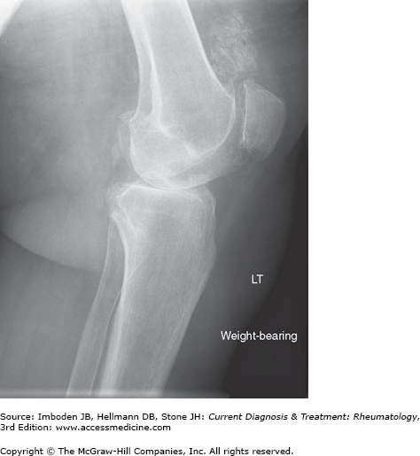
Synovial chondromatosis is a rare tumor-like condition in which metaplastic synovial lesions develop into cartilaginous islands that in turn produce multiple chondroid loose bodies, which eventually calcify. The process is monoarticular and indolent; the typical presentation is chronic swelling of a knee, hip, or shoulder. In the early phases, radiographs may be unremarkable but, in the later stages, calcified loose bodies—sometimes numbering in the hundreds—are visible (Figure 56–1). Synovial chondromatosis is self-limited but can produce painful locking and secondary osteoarthritis. Surgical removal of loose bodies and synovectomy are usually effective.
Arthritis Due to Direct Involvement by Malignancy
- Rapidly growing, eccentric mass associated with a joint, tendon, or tendon sheath in an extremity.
- Deep, radiating pain.
- Characteristic histologic and cytogenetic findings.
Synovial sarcomas typically present as an enlarging mass in an extremity during the third to fifth decades of life. The mass is associated with tendon, tendon sheath, or joint capsule but rarely is truly intra-articular. In 20–40% of cases, plain radiographs of the mass reveal amorphous calcification that points to the diagnosis. MRI can delineate the extent of the lesion. Treatment consists of wide surgical excision. The 5-year survival rates range from 25% to 60%, and tumor size is an important prognostic factor.
Related posts:


Stay updated, free articles. Join our Telegram channel

Full access? Get Clinical Tree


