Revisional surgical intervention for lower limb peripheral nerve injuries is challenging. Patients with intractable neurogenic pain are at increased risk of functional deficits, socioeconomic debilitation, and narcotic abuse.1–5 Nerve injuries have different clinical presentations depending on the degree and presence of neuronal damage. There are different procedures and interventions described in the literature for treating nerve entrapments and neuromas with varying degrees of pain relief. Recently, surgical intervention for more severe nerve injuries trends toward reconstructive procedures such as nerve transposition with relocation, autologous or allograft reconstruction and targeted muscle reinnervation (TMR), and regenerative peripheral nerve interfaces (RPNI).1,3,6,7 In this chapter, the authors discuss the clinical workup and surgical algorithm currently utilized when treating revisional lower extremity nerve entrapment and neuromas.
Nerve Injuries
As described in Chapter 20, the Seddon and Sunderland classification for nerve injuries is the most common one utilized and is important to understand prior to surgical intervention.8,9
A fourth-degree injury forms a neuroma-in-continuity (Figures 21.1 and 21.2). There is disruption present within the endoneurium and perineurium, but the epineurium remains intact.
Figure 21.1Superficial peroneal nerve neuroma-in-continuity.(Photograph courtesy of Dr. Peter Bregman, all rights reserved.)
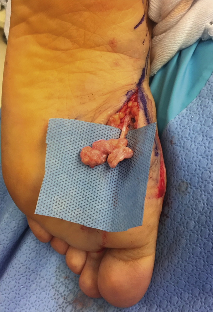
Figure 21.2 Mortons neuroma: Interdigital neuroma-in-continuity within second interspace.(Photograph courtesy of Dr. Peter Bregman, all rights reserved.)
Neurotmesis is associated with a fifth-degree nerve injury and is most commonly seen in fractures, lacerations, crush traumatic injuries, and surgical trauma when the nerve is completely severed and forms an end-neuroma (Figure 21.3). The nerve conduction study shows total absence of nerve conduction velocities and amplitude since there is a total disruption of the nerve.9
Figure 21.3 End or stump neuroma.(Photograph courtesy of Dr. Edgardo Rodriguez-Colazzo, all rights reserved.)
History and Physical Examination
A thorough clinical and bilateral physical examination must be performed prior to further surgical intervention.
History of Past and Present
Regarding the patient’s history of past and present nerve injury, one must consider the following: mechanism of injury (Table 21.1), temporal sequence of pain development, degree of impact and level of dysfunction, and goals of treatment.6
Table 21.1
Mechanism of Injury and Injury Examples That Can Lead to Intraneural Damage
Mechanism of Injury
Examples
Traction
Ankle sprain, fracture, and dislocation/subluxation
Crush
Compartment syndrome
Ischemia
Can occur from extrinsic compression or vascular disease
Systemic disease
Double crush syndromes, diabetes mellitus
Biomechanical
Changes to tarsal tunnel volume due to hindfoot position
Pharmacologic
Chemotherapy, radiation
Providers should also consider the time frame since nerve injury. Even with the correct diagnosis and appropriate surgical intervention, pain can be centralized. Developing an approach with a team, such as a pain management specialist, is encouraged.10
Pearls for Identifying Type of Nerve Injuries:
Although symptoms sometimes vary from person to person, nerve entrapments present with localized or referred symptoms such as11,12:
The physical examination should include evaluation at all possible entrapment sites as there can be multiple sources of neurogenic pain. Whether an entrapment, or neuroma, is present within the lower extremity or the foot, the same principles apply when formulating a diagnosis and treatment plan. For a complete and detailed physical examination and diagnostic studies regarding patients with lower extremity nerve entrapments please refer to Chapter 20 under History and Physical Examination and Diagnostic Studies sections.
Reconstruction and Zone of Injury
In reconstructive surgery, it is imperative to consider the “Zone of Injury,” or the area where the soft tissue has been damaged or previously violated. The zone of injury is divided into 2 categories: nonreconstructible and reconstructible (Table 21.2). In order for a zone of the lower extremity to be considered reconstructible the skin tissue envelope must be healthy and vascularity intact to the nerve bed and extremity. A zone of injury becomes nonreconstructible when the skin tissue envelope is not healthy or is nonmobile, there is scarring, or severe adhesions are present. If there is inadequate vascularity to the zone of injury, it also becomes a nonreconstructible site. If surgical intervention is warranted, the surgeon must consider where, or if, there is a reconstructible site available. The treatment cannot be worse than the disease.
The patient and provider need to thoroughly discuss the patient’s expectations of surgery. The goal of nerve surgery is to provide pain relief, not always improvement of sensation. The patient should expect numbness postoperatively when performing revisional reconstructive procedures, which is essential for patients to understand and the responsibility of the provider to inform the patient.
Surgical Procedures and Algorithm for Nerve Injuries
As mentioned earlier in the chapter, surgical treatments for nerve entrapment and neuromas can be divided into passive/ablative or active/reconstructive procedures.1,3 Below we discuss surgical procedures and provide an algorithm to approach entrapments and neuromas of the lower extremity based on type of nerve injury. Remember, not all patients with a nerve injury require surgical intervention and decisions need to be made on a case-by-case situation.
Depending on the severity of a neuropraxic injury, some patients’ symptoms resolve with conservative management. When conservative management has been exhausted, there are several procedures to consider: decompression with fasciectomy, external neurolysis, internal neurolysis, and perineurolysis. The goal of the surgical treatment is to alleviate the external, or internal, pressure causing damage to the myelin. The myelin will eventually heal and allow the nerve to glide and function without inhibition.13,14
Only gold members can continue reading. Log In or Register to continue


 Postoperative Pain Control and Rehabilitation in Revisional Foot and Ankle Surgery


