Lucian P. Jiga, Zaher Jandali
The Vasculoplastic Approach for Revisional Plastic Surgery of the Diabetic Foot
Introduction
Preoperative Considerations

Clinical Cases
Case 1


Related posts:
 Postoperative Pain Control and Rehabilitation in Revisional Foot and Ankle Surgery
Revisional Pes Planovalgus Deformity Correction
Revision of Failed Osteochondral Lesions of the Talar Dome
Revisional Insertional Achilles Tendon and Haglund Deformity
Revisional Calcaneal Fractures Repair
Revisional Ankle and Tibiotalocalcaneal Arthrodesis
Postoperative Pain Control and Rehabilitation in Revisional Foot and Ankle Surgery
Revisional Pes Planovalgus Deformity Correction
Revision of Failed Osteochondral Lesions of the Talar Dome
Revisional Insertional Achilles Tendon and Haglund Deformity
Revisional Calcaneal Fractures Repair
Revisional Ankle and Tibiotalocalcaneal Arthrodesis
![]()
Stay updated, free articles. Join our Telegram channel

Full access? Get Clinical Tree


The Vasculoplastic Approach for Revisional Plastic Surgery of the Diabetic Foot
Diabetic foot ulcerations (DFUs) in the context of peripheral neuropathy, arterial disease, and foot deformity build up a risky triad that, if not timely addressed, leads to significant tissue loss or even major limb amputation.1 Radical débridement, infection control followed by vascular augmentation, and soft-tissue reconstruction are all instrumental parts of an established vasculoplastic approach to address such limb-threatening conditions with the aim to reduce the overall number of major amputations in the diabetic patient population.2,3
Since its inception over 3 decades ago, the vasculoplastic approach for treatment of recalcitrant limb-threatening DFUs has undergone several major evolutionary milestones. Initially, there was the distal bypass surgery to the vessels of the leg or foot and the autologous venous loops, which, besides improving the vascularity of the affected limb, provided optimal recipient vessels for the free muscle flaps used to cover the soft-tissue defects.4 Major technical breakthroughs in endovascular surgery allowed minimal invasive openings of the distal vascular arches of the foot, providing adequate recipient vessels close to the lesion, which in turn induced a further refinement in the flap choice and aesthetics of the reconstruction (eg, perforator flaps, supra-thin flaps), and represented the next evolution.5 Recently, Hong et al using the supramicrosurgical techniques (vessels with an inner diameter <1 mm) demonstrated the utility perforator vessels, arising from severely calcified main arteries as recipient vessels for free perforator flaps to cover limb-threatening DFUs.6 Such perforator vessels lay always suprafascially, are easily identifiable, and can be approached very fast during surgery while avoiding sometimes complicated dissection (eg, scaring due to previous bypass surgery) to the main trunks in search of an adequate recipient vessel. This new concept has opened an entire new dimension in our approach to ischemic soft-tissue loss in the diabetic population. Supramicrosurgical techniques require, however, unique operative skills and are therefore limited to a handful of reconstructive microsurgeons worldwide.
As the regenerative role of adipose-derived cells receives increasing attention, several groups were able to demonstrate the beneficial effects of autologous lipoaspirates on DFU healing, reporting up to 95% complete wound closure 3 months after treatment.7 Similarly, other groups investigated the effect of synthetic bilayered collagen matrix on intractable DFUs, reporting up to 80% wound healing rates 12 months postoperatively.8 Furthermore, this process necessitates a significant amount of wound healing time, meanwhile patients being bound to open wound therapy and minimal to non–weight-bearing activities.
In contrast, vascular augmentation in the context of radical débridement and infection control followed by microsurgical soft-tissue reconstruction provides a straightforward concept for wound closure in DFUs, rendering fully ambulating patients within short time after treatment initiation. Both autologous fat transfer and synthetic collagen matrix applications have clear beneficial effects on DFU healing; however, these procedures might possibly find indications in wounds of limited dimensions without exposure of vital structures needing preservation and where all other operative treatments have failed.
In this chapter, the authors present their experience to the vasculoplastic approach for diabetic limb salvage procedures, focusing on the role of microsurgery to provide adequate tissue where other measures of wound management or closure have failed.
DFUs represent the consequence of a complex milieu of pathological mechanisms, from foot deformity with loss of physiological cushioning to vascular impairment and neuropathy. Therefore, a multidisciplinary approach including but not limited to orthopedic surgeons, radiologists, vascular surgeons, plastic surgeons, podiatric surgeons, internists, and infectious disease specialists become mandatory to warrant therapy success. Besides the chronically recurring DFUs, another category of diabetic patients with progressing nonhealing wounds is dealing with wound recurrence in spite of previous wound treatments and grafted acute diabetic foot infections in the context of major vascular impairment.
Revisional surgery for a failed plastic reconstruction in the diabetic population will always pose a particular challenge to the plastic surgeon. The presence of scars either from nonhealing wounds or previous incisions as well as nonusable “expired” recipient vessels from previous surgeries makes the clinical decision process particularly difficult. In an effort to simplify the thought process to this particular type of DFUs, the authors have developed an approach algorithm that proved to be very efficient in terms of successful wound closure (Figure 32.1). The following case presentations show our concept in approaching revisional DFU surgery and reconstruction.
A 67-year-old diabetic patient referred to our clinic for intractable progressing ulceration of the right plantar hindfoot with a DFU as the initiating event. The patient had been treated with a total of 8 successive débridements, 2 failed skin grafts, and vacuum-assisted open wound treatment as last option for several weeks before being referred to us.
Initial clinical examination showed a complex soft-tissue defect involving the entire calcaneal region with exposed bone and part of the plantar fascia over the entire otherwise clean wound bed (Figure 32.2). The dorsalis pedis and posterior tibial artery as well as the popliteal artery were pulseless. Wound cultures were negative. Furthermore, the patient had a history of ischemic heart disease, double coronary artery bypass grafting, a cerebrovascular event due to bilateral internal carotid artery stenotic disease treated with open eversion endarterectomy, and a body mass index of 32 kg/m2. In spite of all medical comorbidities, the patient had a good general condition and was ambulatory with off-loading the right foot.
Figure 32.2 Preoperative image of the wound with exposed calcaneus and plantar fascia.
Magnetic resonance imaging angiograph revealed advanced peripheral arterial disease (PAD) with several high-grade stenotic diseases of the superficial femoral artery, tibioperoneal trunk, and proximal peroneal artery (Figure 32.3A). Under these circumstances, the lower extremity was considered to be salvageable.
Figure 32.3 A, Magnetic resonance angiogram showing poor vascular status of the left lower extremity with obstruction of the superficial femoral artery (SFA), tibioperoneal trunk, and proximal peroneal artery (PA). Both lesions were treated by percutaneous transluminal angioplasty (PTA); B, SFA recanalization; C and D, recanalization of the tibioperoneal trunk.
Revascularization was performed first by endovascular opening of the superficial femoral artery, tibioperoneal trunk, and the proximal peroneal artery using percutaneous transluminal angioplasty (PTA) (Figure 32.3B-D). After PTA, the peroneal artery became the only available source vessel in continuity for soft-tissue reconstruction. Preoperative Doppler ultrasound evaluation (18-MHz linear probe) of the perforators arising from the distal peroneal artery revealed at least 2 perforator pedicles with an acceptable location from the proximal wound margin, which could be considered as pedicles for a local perforator flap. Alternatively, the middle third of the peroneal artery was evaluated as possible recipient vessel for a free flap. However, because of the necessary lateral approach for its adequate exposure at this level necessitating resection of the fibula, the long operative time for a free flap, and severe mediocalcosis all along the entire vessel length, it was decided to explore first the perforators and the possibility to harvest a local perforator flap to cover the defect.
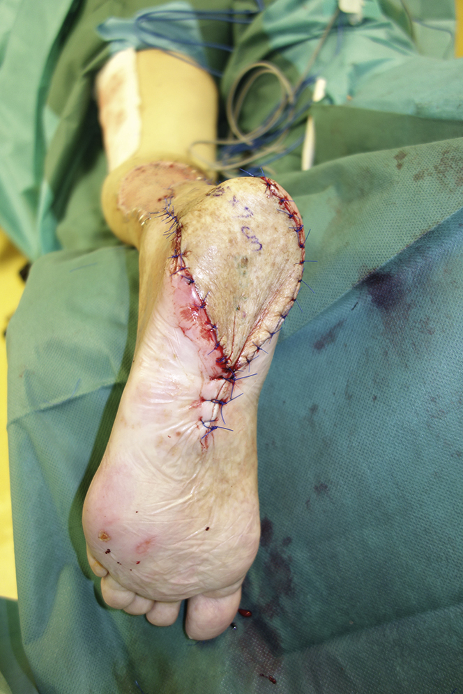
With the patient in prone position, the first exploratory incision was made dorsally approximately 2 cm anterior from the skin projection of the lateral border of the Achilles tendon (Figure 32.4). After incising the fascia, the perforator pedicles were inspected and the one closer to the proximal wound margin was chosen. With the area where this perforator pierces the fascia marked as the pivot point and taking into consideration the width of the defect, the final dimensions of the flap were set and marked. Flap was harvested subfascially from proximal to distal. It is important to free the perforator pedicle of any fascial attachments. Once completely harvested, the flap is turned 180°, thereby achieving a tension-free wound closure (Figure 32.5). When flaps of more than 4 cm width are harvested from the distal lateral shank, the donor site will usually need autogenous or allogeneic skin grafting for an optimal closure as in our patient (Figure 32.6).
Figure 32.5 The propeller flap rotated into the defect.A tension-free closure could be achieved.
Figure 32.6 Closure of the donor site using autologous split-thickness skin grafting.
As in any flap surgery, avoiding pressure on the flap in the early postoperative period is instrumental to warrant optimal healing. Thus, when reconstructing defects on the plantar aspect of the foot or dorsal aspect of the leg, our patients will receive a minimum of 5 days of an external fixator application, which provides excellent means for complete isolation of the operated area by attaching the foot in a bed-mounted extension device (Figure 32.7). Additionally, as we have found it beneficial, all our flap patients will receive on the flap within the fifth postoperative day warm air therapy (38 °C) using an air circulated blanket. Adequate pain medication and antibiotic coverage round up our postoperative care protocol.
