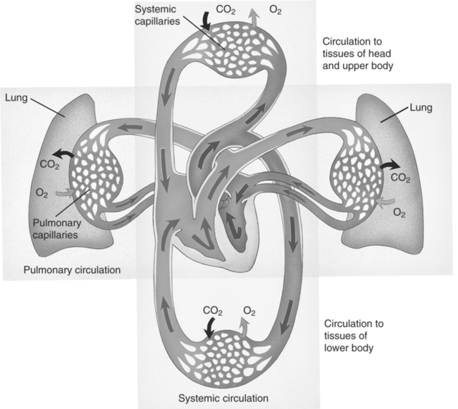
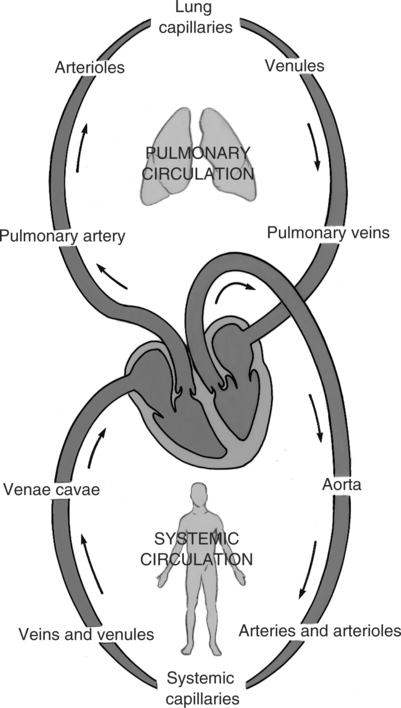
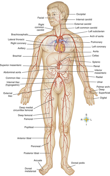
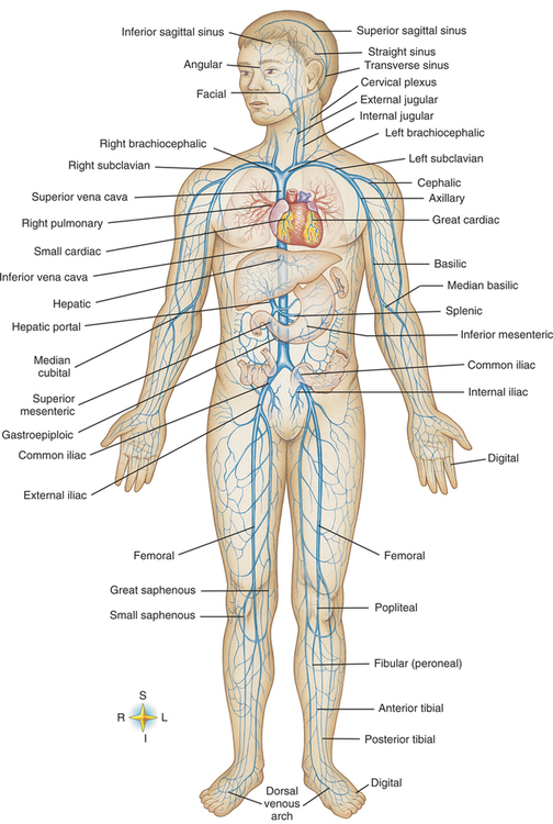
Review of Anatomy and Physiology

Chapter 2


Review of Anatomy and Physiology


Key Terms


aorta


the main trunk arterial vessel (artery) that leaves the left ventricle and carries blood to systemic arteries.


aortic valve


heart valve between left ventricle and aorta.


arterioles


very small arteries at the distal end of the arterial network that carry blood to the capillaries.


artery


blood vessel that carries blood away from the heart that in most cases is oxygenated.


capillaries


minute blood vessels that connect the smallest arterioles to the smallest venules. The walls of capillaries are a single layer of epithelial cells enabling blood and tissue fluid the ability to exchange various substances (gases, fluids, electrolytes, salts, and nutrients).


diffusion


movement of particles or molecules from an area of higher concentration in fluids to one of lower concentration to achieve more equal distribution of particles/molecules in the fluid or liquid.


extracellular


outside the cell.


filtration


process in which particles are removed from solution by passing the liquid through a membrane or filter.


homeostasis


state of dynamic balance in the body.


hydrostatic pressure


pressure exerted on a liquid.


inferior vena cava


large vein that collects blood from parts of the body below (inferior to) the heart for return to right atrium.


interstitial


pertaining to space between cells.


interstitial fluids


extracellular fluid in the spaces between body cells providing a substantial portion of liquid environment of the body.


intracellular


within the cell.


intravascular fluids


fluids within the blood vessels.


left atrium


left upper chamber of the heart; receives blood from the pulmonary veins.


left ventricle


left lower chamber of the heart; receives blood from the left atrium.


mitral or bicuspid valve


valve between the left atrium and left ventricle.


osmosis


passage of solvent (fluid) through semipermeable membrane that separates different concentrations of solutions. Movement is from an area where there is a lower concentration of solute into an area where there is an area of higher concentration of solute to equalize the concentration of the two solutions.


plasma


serum, proteins, and chemical substances found in the aqueous portion of blood.


pulmonary circulation


circulation of blood through the lungs for exchange of oxygen and carbon dioxide.


pulmonary (semilunar) valve


valve between the right ventricle and the pulmonary artery.


right atrium


right upper chamber of the heart; receives blood returning to the heart from the inferior and superior vena cava.


right ventricle


right lower chamber of the heart; receives blood from the right atrium.


solute


substance dissolved in a solution.


solvent


solution holding the solute.


superior vena cava


large vein that collects blood from parts of the body above (superior to) the heart for its return to the right atrium.


total parenteral nutrition (TPN)


nutritional support via central access IV therapy to provide glucose, proteins, vitamins, electrolytes, sometimes fats, and so on to maintain the body’s growth, development, and tissue repair.


tricuspid valve


valve between the right atrium and right ventricle.


veins


vessels that return blood toward the heart.


venipunctures


puncture of a vein for any purpose; related to IV therapy, puncture of the vein to place an administration device for infusion of IV fluids.


venules


very small veins that carry blood from the capillaries to the veins.


The health care clinician responsible for intravenous (IV) therapy must be cognizant of many aspects of anatomy and physiology, including cell structure; circulatory, lymphatic, and nervous systems; and blood and its components. Reviewing the anatomy of the general cardiovascular system stimulates an awareness of its structures, especially the peripheral vessels, as used with IV therapy. Reassessing the peripheral vascular system and its structures allows the clinician to select an appropriate site for the initiation of IV therapy. Studying the physiology of the circulatory system, including the cardiovascular and peripheral vascular system, helps the clinician to understand the flow of the blood through the body.


Intravenous therapy is not totally dependent on the circulatory system, but includes the relationship with other body systems. Reviewing the involvement of lymph and the lymphatic system helps in understanding the circulation of lymphatic fluid and its relationship to blood in the body. Becoming aware of the nerve structures in the area surrounding the peripheral blood vessels is essential to prevent injury to the nerves at the venipuncture site and to reduce patient pain. Components and properties of blood are also factors in successful IV therapy. Additionally, the clinician should review the role of water, electrolytes, osmolarity, and pH in homeostasis of the individual. Intracellular fluids (ICF), extracellular fluids (ECF), interstitial fluids, and the pH of the fluids (such as isotonic fluids, hypotonic fluids, and hypertonic fluids) all are considered for IV therapy and patient safety.


HOMEOSTASIS


Homeostasis is defined as the relative constancy of the internal environment of the human body that is maintained by adaptive responses to promote health and survival. The steady state is maintained through feedback and various sensual mechanisms that provide organs with the needed responses to maintain this internal balance essential for survival. Balance of the body’s internal environment includes balancing of body fluids and chemical substances, some of which include electrolytes that are dissolved in body fluids. This dynamic equilibrium between the fluids and electrolytes involves the delicate balance needed to maintain homeostasis and support processes necessary to maintain life. IV therapy is used to maintain homeostasis for those people who for whatever reason have the need for replacement of fluids or electrolytes as well as for medication therapy and blood product replacement.


Body cells are the fundamental functioning unit of the human body. For cells to be able to perform the necessary tasks for maintenance of life, a stable environment, or homeostasis, is necessary. The stability is based on the constant supply of nutrients for the body and the continuous excretion of wastes from metabolism through the urinary, gastrointestinal, or integumentary systems, with some small amounts being lost through respiration. The body fluids lost by removal of body waste must be replaced for homeostasis to be maintained.


Water, accounting for approximately 60% to 75% of total body weight, is the single largest constituent of the body’s mass and is essentialto life. Fat cells contain less water, making the percentage of body water dependent on the fat distribution of the person. Age, gender, ethnic origin, and weight also are factors that influence the amount and distribution of body fluids.


Continually moving in the body, water is given different names in various locations, such as intracellular (ICF), extracellular (ECF), plasma, lymph, and interstitial fluid. Homeostasis depends on fluid and electrolyte intake and physiologic factors, disease processes, external factors, and pharmacologic interventions. Electrolytes for homeostasis are dissolved in the blood plasma, a body solvent, for transport throughout the body. Body fluids are continually exchanged in the intracellular and extracellular sites, such as interstitial spaces and plasma. For a person to remain in fluid homeostasis, that person must maintain an approximately equal intake to output of fluids.


Many of the substances normally found in the body exist in solution, usually aqueous base. The water or fluid is found in different compartments of the body. Intracellular fluid is fluid contained within the cells. Extracellular fluid is found in the spaces between the cells (interstitial space) and in the vascular network as plasma. Fluids have the ability to shift from one area to another, depending on the concentration of electrolytes and proteins in the areas. Fluids will move from an area of lowest concentration of solutes to that of the greatest concentration of solutes in a solvent to bring the solution closer to equilibrium of concentration. Examples of consequences of fluid shift include dehydration from vomiting and diarrhea with resulting greater concentration of electrolytes in extracellular spaces. Conversely, fluid retention in the extracellular spaces is treated by restricting sodium and fluid volumes and by drug therapy.


An additional factor in homeostasis or fluid balance is the health status of the person. Dehydration can be caused by extended periods of vomiting or diarrhea, exposure to extreme heat, and reduction of intake of oral fluids. Conditions such as impaired cardiac, renal, or liver function also play an important role in homeostasis. Trauma or surgical procedures may also cause a fluid and/or electrolyte imbalance.


CIRCULATORY SYSTEM


A review of the general circulatory system is vital for patient safety with IV therapy. The anticipation is that this review will provide a recollection of knowledge previously gained.


The heart, located in the thoracic cavity, is situated in the middle of the mediastinal region between the two lungs. It lies behind the sternum and in front of the spinal column. Although the size of the heart varies in each individual, the normal heart, the size of a closed fist, acts as a muscular pump that circulates blood to all tissues of the body.


Route of Blood Flow


Refer to Figures 2-1 and 2-2 for primary routes of the circulation system. The chamber of the heart first receiving carbon dioxide (CO2)-laden blood from the superior and inferior vena cava is the right atrium. The blood, moving primarily by gravity as the valves open, travels through the tricuspid valve into the right ventricle. The walls of the atria are thinner than the walls of the ventricles because they do not have to pump the blood into the next area against gravity and to more distant areas of the body as the ventricles do. The right ventricle pumps blood through the pulmonary (semilunar) valve into the pulmonary artery and then into the lungs, or through pulmonary circulation. The blood travels through pulmonary circulation where gases CO2 and oxygen (O2) are exchanged at the cellular level of the alveoli of the lungs. The oxygen-laden blood leaves the pulmonary circulation via the pulmonary veins to return to the heart into the left atrium. The blood then passes through the mitral or bicuspid valve into the left ventricle. Here, the forceful contraction of the cardiac muscle accelerates the blood through the aortic valve into the aorta and into the cardiac and general body or systemic circulation.


The oxygenated blood travels through the systemic circulation to the body via the aorta, which branches to form arteries to the myocardium (or coronary circulation through coronary arteries), brain (through the carotid and cerebral arteries), upper limbs (through brachiocephalic and subclavian arteries), and trunk and lower limbs (via thoracic aorta and femoral arteries) (Figure 2-3 and color insert). As the aorta descends into the body, it divides into arteries to supply circulation to the major organ systems and to the peripheral regions of the body. As the arteries branch out, they gradually become smaller and are termed arterioles. At the terminal branches of the arterioles, capillaries with permeable membranes are formed to allow transfer of nutrients and oxygen into the tissues and absorption of CO2 and metabolic waste products for return to the heart. The distal end of capillaries become very small veins, called venules, and as they progress back to the heart, they turn into progressively larger veins (Figure 2-4 and color insert). The veins terminate in the superior and inferior vena cava to complete the circulatory circuit as they empty deoxygenated blood into the right atrium of the heart.


Arteries carry blood away from the heart, and veins return blood to the heart. Systemic arteries are responsible for transporting oxygen-rich blood from the heart to the tissues throughout the body. The only arteries that do not carry oxygen-laden blood are the pulmonary arteries that carry the deoxygenated blood away from the heart to the lungs. This blood, laden with CO2, will be transported through the pulmonary arteries for exchange of CO2 and O2 in the lungs. The pulmonary veins (the only veins that carry oxygenated blood) return the oxygen-rich blood back to the left side of the heart for circulation to the body by systemic circulation.


The oxygen-laden blood travels through the arteries to the arterioles and then to the capillaries, where the thin one-celled walls of the capillaries facilitate exchange of gases, nutrients, and solutes into the tissues. The capillaries, residing in tissues, receive waste products of nutrients and CO2 following metabolism and return these to the blood. The capillaries progress into venules and then veins for the blood to eventually return to the heart via the inferior and superior vena cavae.


Fluid containing solutes leaves the capillaries to surround tissue cells as extracellular fluid.

Stay updated, free articles. Join our Telegram channel

Aug 10, 2016 | Posted by in PHYSICAL MEDICINE & REHABILITATION | Comments Off on Review of Anatomy and Physiology

Full access? Get Clinical Tree

Get Clinical Tree app for offline access