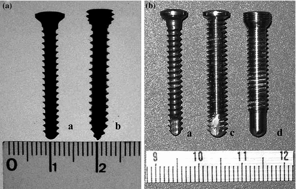
Fig. 4.1
Grammont reverse
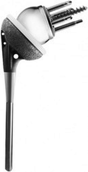
Fig. 4.2
DJO monoblock reverse
Scapula-Sided Biomechanics
A great deal of effort has been spent on trying to understand scapula-sided problems in RSA. From baseplate failures to scapular notching and how the position of the glenosphere can affect ROM, being able to understand how all these factors interact with each other remains of vital importance to our institution.
Baseplate Failures and What Can Be Done to Lessen Their Incidence
Baseplate failures were a concern early on with the RSP. Because of the more lateral nature of the COR of the glenosphere, it was believed that the increased torsional moments seen by this implant would eventually cause this implant to fail. Although there were a limited number of initial failures of this device, through the research presented here, it was concluded that the main mode of failure involved micromotion between the non-locking peripheral screws and the baseplate. The following study helped elucidate this problem and guided future studies into this problem.
Scanning Electron Microscopy of Failed RSP Baseplates [16]
The purpose of this study was to identify possible causes of baseplate failure in retrieved RSP baseplates. Two failed baseplates were scanned using a scanning electron microscope (SEM) to determine possible modes of failure and identify whether bony ingrowth had occurred. Results showed striations at the fractured central screw that were characteristic of cyclic fatigue failure (Fig. 4.3a and b). Additionally, minimal bony ingrowth was identified on the porous underside of the baseplate. These findings suggested a gradual failure of the central screw due to the failure to achieve bony ingrowth into the porous underside of the baseplate. This failure to achieve ingrowth was ultimately attributed to macromotion between the baseplate and the peripheral non-locked screws. This study ultimately helped guide our research to identify possible solutions to this problem.
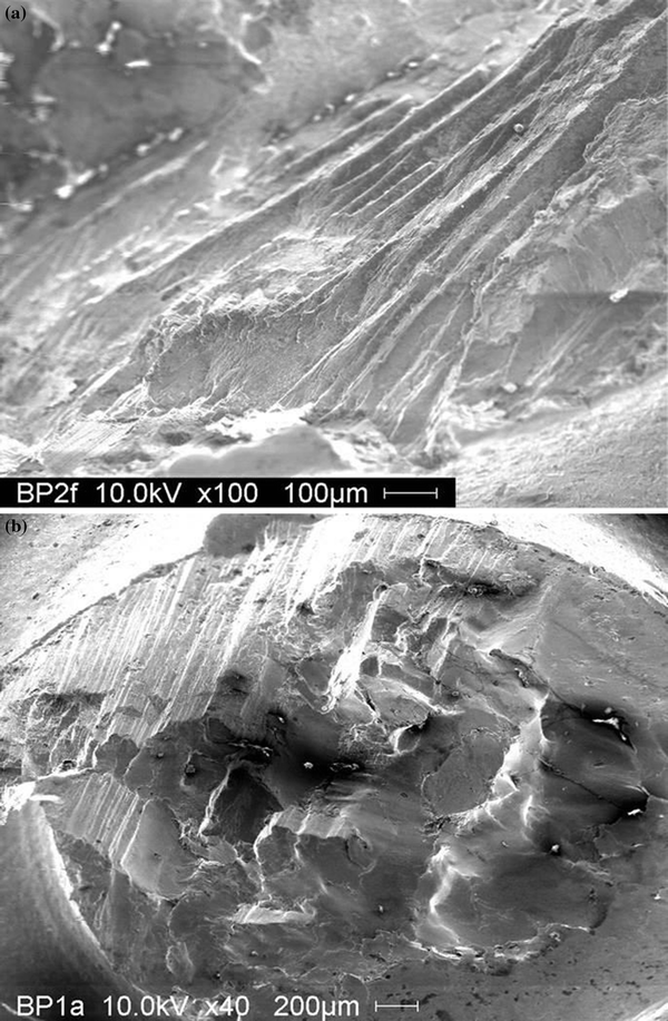
Fig. 4.3
SEM of baseplate failure
Baseplate Fixation
Once possible causes for baseplate failure were identified, focus turned toward understanding the factors involved in baseplate fixation. To this end, the following 4 studies were undertaken involving multiple institutions with their ultimate goal of improving patient outcomes in RSA.
Compressive Strength of Central Screw [17]
The purpose of this study was to determine the compressive force present between the baseplate and the glenoid for two different types of baseplates: (1) a Grammont style with a smooth central post and (2) an RSP style with a 6.5-mm integrated central cancellous-type screw. Both devices were implanted to manufacturer’s specifications including peripheral screws tightened to 60 in./lb. Film-type force transducers were positioned between the baseplates and a bone analog to measure compressive force. Results showed a tenfold increase in compressive force when a central cancellous-type screw was used instead of a smooth central peg (2000 N vs. 200 N, respectively). This study showed the benefit of having a baseplate with a central cancellous-type screw since it provided a tenfold increase in compressive force over a smooth central post secured only by peripheral screws.
Maximum Load to Failure of Central Baseplate Fixation [18]
The purpose of the study was to determine resistance to shear force for two different types of baseplates: (1) a Grammont style with a smooth central post and (2) an RSP style with a 6.5-mm integrated central cancellous-type screw. Both devices were implanted to manufacturer’s specifications, but without the use of peripheral screws. A shear force of 756 N was applied to the rim of each baseplate at a rate of 150 N/s until the yield strength of the bone analog was reached (Fig. 4.4). Results showed an almost threefold increase in resistance to shear force when a central cancellous-type screw was used instead of a smooth central peg (631 N vs. 269 N, respectively). This study added to the evidence for having a baseplate with a central cancellous-type screw.
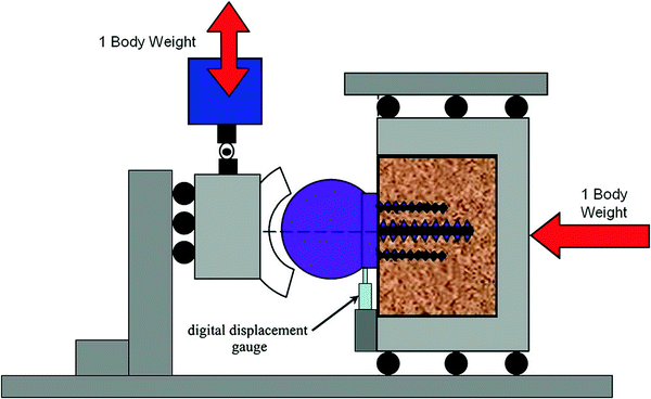
Fig. 4.4
Study setup showing compressive force applied to glenosphere. Used with permission from Virani et al. [36]
Baseplate Micromotion Using Locked and Non-Locked Peripheral Screws [19]
The purpose of this study was to quantify baseplate micromotion when 3.5-mm non-locking and 4.5-mm locked peripheral screws were used for two different types of baseplates when implanted in a bone analog: (1) a Grammont style with a smooth central post and (2) an RSP style with a 6.5-mm integrated central cancellous-type screw (Fig. 4.5). Both devices were implanted to manufacturer’s specifications. The Grammont-style baseplate was implanted with two 3.5-mm non-locked and two 4.5-mm locked peripheral screws, while the RSP was implanted using only 3.5-mm non-locked peripheral screws. A shear force of 756 N was applied to the rim of each baseplate at a rate of 150 N/s until the yield strength of the bone analog was reached. Results showed a significantly higher resistance to shear force when a central cancellous-type screw was used instead of a smooth central peg (367 ± 23 µm vs. 310 ± 20 µm, respectively). This study showed the benefit of using a central 6.5-mm compressive cancellous screw compared to a central post, even when using compressive peripheral screws combined with locked peripheral screws.
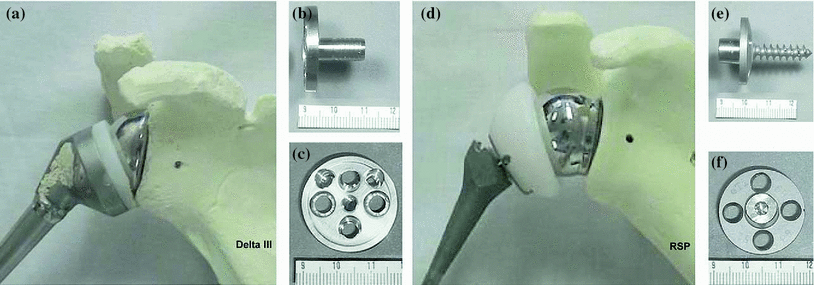
Fig. 4.5
Illustrations of Grammont-style implant (A-C) and DJO implant (D-F). Used with permission from [20]
Glenoid Component Fixation in Three Different RSA Glenosphere Designs [20]
The purpose for this study was twofold:
1.
Compare glenoid component micromotion in three different RSA designs: (a) a Grammont style with a smooth central post and a 36-mm glenosphere with COR at the glenoid, (b) an RSP style with a 6.5-mm integrated central cancellous-type screw and a 32-mm glenosphere with a COR 10 mm away from the glenoid, and (c) an RSP style with a 6.5-mm integrated central cancellous-type screw and a 32-mm glenosphere with a COR 6 mm away from the glenoid (Fig. 4.6). All devices were implanted into a bone analog using manufacturer’s specifications. The Grammont-style baseplate was implanted with two 3.5-mm non-locked and two 3.5-mm locked peripheral screws, while the RSPs was implanted using the following peripheral screw combinations: (a) four 3.5-mm non-locked peripheral screws, (b) four 5.0-mm non-locked peripheral screws, and (c) four 5.0-mm locked peripheral screws (Fig. 4.7). A shear force of 756 N was applied to the rim of each baseplate at a rate of 150 N/s until the yield strength of the bone analog was reached. Results showed a higher resistance to shear force in the Grammont glenoid components when compared to the RSP glenoid components, although all micromotion stayed below 150 µm. When comparing only the RSP designs; the 5.0-mm locked peripheral screws had less mean micromotion than either the 5.0-mm (p = 0.016) or 3.5-mm (p = 0.067 non-locked screws.
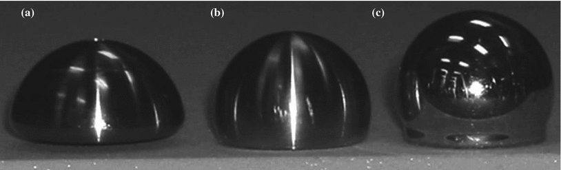
Fig. 4.6
The different glenospheres used in this study: a Grammont, b RSP Reduced—6 mm offset and c RSP Neutral—10 mm offset. Used with permission from [20]
2.
Compare the effect of lateralization on glenoid component micromotion using two different types of peripheral screws: (a) 3.5-mm non-locked peripheral screws and (b) 5.0-mm locked peripheral screws. An offset gauge device was used to apply cyclic shear forces (±756 N) to varying lateral offsets (from 2 to 30 mm). Results showed a positive linear correlation between glenoid component micromotion and lateral offset when either of the two types of peripheral screws were used. Additionally, glenoid components fixed with 5.0-mm locked screws showed a 29 % decrease in micromotion when compared to the 3.5-mm non-locked screws.
This study, as with the previous study, showed the benefit of using 5.0-mm locked screws over 3.5-mm non-locked screws. The information presented in this section helped modify surgical technique by recommending the use of 5.0-mm locked peripheral screws when implanting an RSP baseplate.
Baseplate Position
Although baseplate failures have been mostly addressed by the use of 5.0-mm locked screws in place of the 3.5-mm non-locking screws, the few failures that do occur remain a concern with the RSP design. Observations by the design surgeon guided new biomechanical studies into how the position of the baseplate might affect the survivability of the baseplate. The following studies aided in the understanding of how altering design parameters and surgical techniques could affect ROM, glenoid forces, and baseplate displacement.
The Effects of Baseplate Tilt on Glenoid Forces—a Biomechanical Model [21]
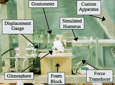
The purpose of this study was to quantify the effects of baseplate tilt (15° superior tilt, 0° neutral tilt, and 15° inferior tilt) on baseplate micromotion and compressive forces seen by the glenoid. An apparatus was developed to simulate 60° of glenohumeral abduction in the scapular plane. Superior and inferior baseplate micromotion was measured via a linear voltage displacement transducer (LVDT), and glenoid forces were measured by transducers that were placed underneath the baseplate (between the baseplate and a bone analog) in the superior and inferior positions (Fig. 4.8). Results showed less overall micromotion and more evenly spread compressive forces when the baseplate was positioned with a 15° inferior tilt. The results from this study correlated well with what was being seen clinically.
The Effects of Baseplate Tilt on Glenoid Forces—a Finite Element Model [22]
The purpose of this study was to compare the effects of inferior tilt (0°, 3°, 6°, 9°, 12° and 15°) on strain between RSP and Grammont-style baseplates. A finite element model was developed to simulate the RSP and Grammont-style devices in a bone analog substrate (Fig. 4.9). Results showed greater baseplate motion in the superior and inferior directions with the Grammont-style device when compared to the RSP design. Additionally, increasing the inferior tilt of the baseplate in either design showed a decrease in overall motion. The results from this study corroborated the evidence found in the previous biomechanical study and ultimately helped to alter clinical practice by advising surgeons to implant the baseplate with up to a 15° inferior tilt.
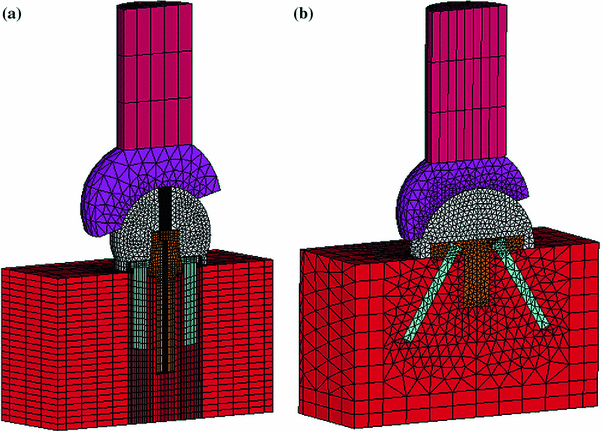
Fig. 4.9
Finite element model showing a DJO implant and b Grammont-style implant. Used with permission from Virani et al. [36]
The Effect of Varying Glenosphere COR on Baseplate Fixation [23, 24]
The purpose of this study was to compare the effects of varying glenosphere COR on (1) baseplate micromotion in two separate studies, (a) biomechanical and (b) finite element model, and (2) reaction moments from a numerical model. Seven different glenospheres (RSP: 32, 36, and 40 mm neutral and 32 mm −4, 36 mm −4 and 40 mm −4; Grammont: 36 mm) with five different CORs (0 mm [40 mm −4], 1 mm [36 mm—Grammont], 2 mm [36 mm −4], 4 mm [40 mm neutral], 6 mm [36 mm neutral and 36 mm −4], and 10 mm [32 neutral]) were tested.
(a)
Biomechanical Model
The testing apparatus used by Harman et al. was also used in this study (Fig. 4.10). The baseplates were implanted into a bone analog using either 5.0-mm locked screws (RSP) or 3.5-mm non-locking and 3.5-mm locking screws (Grammont). A shear force of 756 N was applied to the rim of each baseplate at a rate of 150 N/s until the yield strength of the bone analog was reached and subsequent micromotion was measured.
Fig. 4.10
Testing setup
(b)
Finite Element Model
A finite element model was created to simulate the biomechanical testing performed in (a) above (Fig. 4.11). It included multiple coefficient of frictions between the glenosphere and humeral socket.
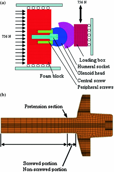
Fig. 4.11
Finite element model showing a a testing setup analogous to Harman et al. 2005c and b boundary conditions for RSP baseplate
(c)
Analytical Model
An equation was developed to predict the effect of changing the coefficient of friction and the COR on the moment applied to the baseplate.
The results from the analytical and finite models showed good agreement with both showing 30 µm or less variability in baseplate micromotion by varying glenosphere designs. The analytical model showed a general increase in moments at the baseplate as both coefficient of friction and COR offset were increased. These results showed minimal differences in motion between all the designs of glenosphere, as long as good initial glenoid fixation was achieved.
Glenohumeral Morphology
Implantation of an RSA baseplate involves taking into consideration multiple anatomical parameters. The knowledge of how these different parameters can affect placement is crucial to the clinician. The following studies were undertaken to help the clinician navigate these intricacies and help improve patient outcomes.
Glenoid Morphology in Reverse Shoulder Arthroplasty: Classification and Surgical Implications
The purpose of this study was to classify glenoid morphology and examine how variances in this morphology can affect the fixation of glenoid components. The morphology of 216 glenoids was divided into normal and abnormal groups according to six anatomic parameters [height, width, version, inclination, distance from coracoid to the glenoid (C-G), and distance from the acromion to the glenoid (A-G)] and 2 surgical parameters (central/standard centerline and spine centerline for central axis device fixation). The abnormal groups were further subdivided by erosion site (posterior, superior, global, and anterior) (Fig. 4.12). Results showed a decrease in the standard centerline distance in abnormal glenoids (19.6 ± 9.1 mm) versus normal glenoids (28.6 ± 4.1 mm, p < 0.0001). Conversely, the spine centerline showed a larger distance in the abnormal glenoids (34.9 ± 17.0 mm). Additionally, peripheral screw placement was reduced by 42 % in abnormal glenoids, with the placement limited to the anterior and inferior quadrants. This study showed the significant effect that abnormal glenoid morphology had on both baseplate and peripheral screw placement, thus altering surgical technique.
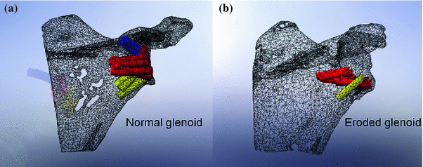
Fig. 4.12
Illustration of a normal glenoid and b abnormal glenoid
Kinematic Impact of Size on the Existing Glenohumeral Joint in Patients Undergoing Reverse Shoulder Arthroplasty [25]
The purpose of this study was to (1) quantify anatomic measurements and glenohumeral relationships and determine whether they change linearly based on patient size, and (2) determine how patient shoulder prosthetic size mismatch can affect soft tissue strain and the middle deltoid moment arm. The anatomic measurements and glenohumeral relationships of 92 patients undergoing RSA, without bony deformities, were evaluated using 3D CT reconstructions. The anatomic measurements were as follows: coronal plane head diameter, thickness of humeral head, neck-shaft angle, articular arc angle, superior humeral head to greater tuberosity distance, lateral humeral contact point to greater tuberosity distance, axial plane head diameter, upper arm length, glenoid height, superior glenoid width, and inferior glenoid width (Fig. 4.13). The glenohumeral relationships were as follows: lateral distance to greater tuberosity, lateral distance of greater tuberosity to acromion, superior distance of greater tuberosity to acromion, lateral distance to coronal head center, lateral distance to axial head center, superior subacromial space, and moment arm. Results showed that humeral head size, glenoid width, lateral offset, and moment arm all independently increased linearly (r2 ≥ 0.92), but the rate of increase varied (range from 0.59 to 1.92). Glenoid height, coronal humeral head diameter, and gender predicted greater tuberosity position within 1.09 ± 0.84 mm of actual position in 90 % of the studied population. Results also showed that patient shoulder prosthetic size mismatch can have an adverse effect on soft tissue strain and middle deltoid moment arm. Strains increased from 3 to 7 % when a small shoulder was implanted with a small implant versus a large implant, respectively. Moment arms were increased by 35 % when a small shoulder was implanted with a large implant, while the moment arm of a large shoulder with a small implant only increased the moment arm by 25 %. This study showed that differences in shoulder measurements and glenohumeral relationships can help group patients by size, sex, and certain morphological measurements. The clinician should be cognizant of choosing the correct size implants based on patient size and should avoid large deviations in non-anatomic reconstructions.
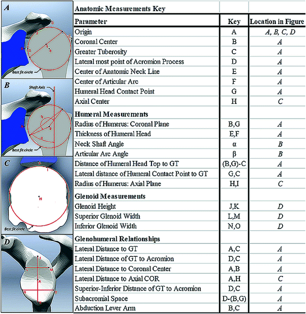
Fig. 4.13
 Intact Rotator Cuff
Origins of Reverse Shoulder Arthroplasty and Common Misconceptions
1 History of Reverse Shoulder Arthroplasty
6 Preoperative Work-up and Surgical Approaches for Reverse Shoulder Arthroplasty
23 Debate: Patient-Specific Instrumentation Is Necessary versus It Is Not Necessary in All Cases
28 The Use of Convertible Glenoid Component Makes Revision to Reverse Total Shoulder Arthroplasty Easier
Intact Rotator Cuff
Origins of Reverse Shoulder Arthroplasty and Common Misconceptions
1 History of Reverse Shoulder Arthroplasty
6 Preoperative Work-up and Surgical Approaches for Reverse Shoulder Arthroplasty
23 Debate: Patient-Specific Instrumentation Is Necessary versus It Is Not Necessary in All Cases
28 The Use of Convertible Glenoid Component Makes Revision to Reverse Total Shoulder Arthroplasty Easier


Illustration of measurements performed in this study. Used with permission from [25]
Related posts:
Intact Rotator Cuff
Origins of Reverse Shoulder Arthroplasty and Common Misconceptions
1 History of Reverse Shoulder Arthroplasty
6 Preoperative Work-up and Surgical Approaches for Reverse Shoulder Arthroplasty
23 Debate: Patient-Specific Instrumentation Is Necessary versus It Is Not Necessary in All Cases
28 The Use of Convertible Glenoid Component Makes Revision to Reverse Total Shoulder Arthroplasty Easier
Stay updated, free articles. Join our Telegram channel
Full access? Get Clinical Tree
