CHAPTER 42 Results and Complications
The results of revision shoulder arthroplasty are as variable as the indications for which it is performed. In general, outcomes after revision shoulder arthroplasty are less satisfactory than those after primary shoulder arthroplasty. Because of the paucity of results of revision shoulder arthroplasty reported in the literature, this chapter reports the results of revision shoulder arthroplasty by drawing from our own experience. Additionally, the most frequent complications and their treatment are outlined.
RESULTS
The results of revision shoulder arthroplasty are hard to examine because of the diversity of indications for which revision is performed. A simple indication such as converting a hemiarthroplasty to a total shoulder arthroplasty for symptomatic glenoid erosion would logically yield a better outcome than would implantation of a revision shoulder arthroplasty for a chronic infection after multiple irrigation and débridement sessions. Unfortunately, the relative rarity of revision shoulder arthroplasty prevents definitive conclusions regarding outcomes. Table 42-1 details the results of revision shoulder arthroplasty from our prospective database initiated in 2003. This table expresses the results in terms of active mobility; patient satisfaction; the Constant score, a shoulder-specific outcomes device incorporating pain, mobility, activity, and strength; and the age- and gender-adjusted Constant score.1,2
INTRAOPERATIVE COMPLICATIONS
Humerus
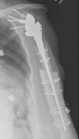
Intraoperative complications involving the humerus are common. The most frequent humeral complication is iatrogenic fracture, which usually occurs during an overly aggressive dislocation maneuver without previous adequate soft tissue release or during extraction of a well-fixed humeral stem. Patients with osteopenia and those with severe preoperative stiffness are most at risk for this complication. These fractures may occur at the humeral diaphysis or proximally and involve the tuberosities. Fractures involving the diaphysis should be reduced and a long-stem humeral implant placed. Allograft struts and cerclage cables may be added in patients with severe osteopenia (Fig. 42-1).
Neurovascular Structures
Catastrophic injury to the neurovascular structures around the shoulder is rare during revision shoulder arthroplasty. The neural structures most at risk during revision shoulder arthroplasty are the axillary and musculocutaneous nerves. If a humeral osteotomy is performed or if revision surgery is performed for a periprosthetic fracture, the radial nerve is also at risk. Nerve injury during revision shoulder arthroplasty can occur as a neuropraxic stretch injury or as a transection injury. Neuropraxic injury caused by stretch most commonly involves the axillary nerve but can involve any nerves within the brachial plexus. Care should be taken when positioning the patient to maintain the cervical spine in neutral alignment to avoid a stretch injury to the brachial plexus. When treating a periprosthetic fracture with revision arthroplasty or when a humeral osteotomy is anticipated for extraction of the humeral stem, the radial nerve should be carefully exposed to ensure its protection. Careful exposure of the radial nerve often results in transient neuropraxia. Patient education preoperatively is of paramount importance in dealing with neuropraxia inasmuch as patients are much more accepting if they have heard about the possibility of this complication before surgery. Axillary and radial nerve neuropraxia is treated by observation, with most patients recovering by 3 to 4 months postoperatively.


