CHAPTER 25 Preoperative Planning and Imaging
Reintroduction of the reverse-design prosthesis has allowed surgeons to treat complicated shoulder pathology for which no good solution existed before availability of this implant. The severity and diversity of shoulder pathology treatable with a reverse prosthesis make preoperative planning even more important in these cases than with primary unconstrained shoulder arthroplasty. Candidates for a reverse prosthesis may include patients with substantial proximal humeral or glenoid bone loss, or both. Because of the complex nature of many of these cases, preoperative planning should be done well in advance of the surgical procedure and not be an afterthought the morning of surgery. As with unconstrained shoulder arthroplasty, preoperative planning for reverse shoulder arthroplasty requires that the surgeon review the patient’s clinical history and physical examination, radiographs, and secondary imaging studies. This chapter outlines our approach to preoperative planning for reverse shoulder arthroplasty.
CLINICAL HISTORY AND EXAMINATION
The same type of clinical history and physical examination is used for candidates for reverse shoulder arthroplasty as for candidates for unconstrained shoulder arthroplasty (see Chapter 7). Because most candidates for reverse shoulder arthroplasty have a compromised rotator cuff, it is important to obtain a detailed history of the patient’s complaints (pain only; weakness only; pain and weakness; pain, weakness, and stiffness).
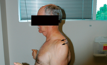
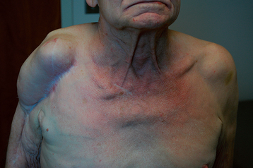
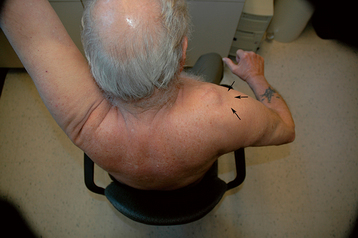
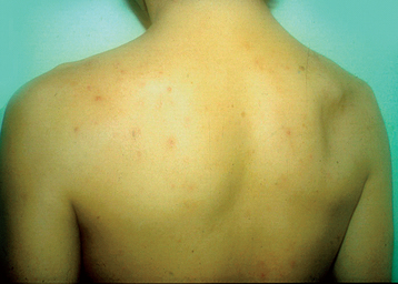
All of our patients undergo a thorough shoulder examination, much of which is detailed in Chapter 7. The visual appearance of the shoulder yields useful information in candidates for reverse shoulder arthroplasty. The presence and location of surgical scars are noted (Fig. 25-1). In thin patients, anterosuperior escape of the humeral head caused by anterior superior rotator cuff deficiency may be obvious (Fig. 25-2) or may be noted only when the patient attempts arm elevation or abduction. Special attention should be paid to the condition of the deltoid, especially if it has previously been surgically violated (Fig. 25-3). The condition of the deltoid is best evaluated by asking the patient to push against your hand to observe an isometric contraction. Any areas of deltoid origin that may not have healed to the acromion after a previous operation should be noted. Atrophy of the supraspinatus and infraspinatus should be noted as well (Fig. 25-4).
Both active and passive mobility is recorded, as detailed in Chapter 7. The presence of glenohumeral crepitus with motion is recorded, as is any discrepancy in active and passive mobility. Special attention should be paid to evaluation of the deltoid muscle. If deltoid contractility appears to be compromised, additional evaluation with electromyography and nerve conduction studies should be performed before further consideration for implantation of a reverse prosthesis.
Rotator cuff examination consists of testing each tendon of the rotator cuff by isolating it as much as possible. Jobe’s test is used to test supraspinatus integrity.1 The infraspinatus is tested by the external rotation lag sign and external rotation strength with the arm at the side.2 The horn blower’s sign is used to test the teres minor.3 The subscapularis is tested with the belly press test and, when mobility allows, the lift-off test.4 Performance of the various rotator cuff tests is depicted in Chapter 7.
The results of the clinical history and examination are documented in the patient’s chart and reviewed well in advance of surgery as part of preoperative planning. Findings are discussed with the patient in terms of postoperative outcome and patient expectations. For example, it is important for patients without posterior rotator cuff function (no infraspinatus or teres minor) to understand that the operation will not restore their ability to actively externally rotate the arm.
Stay updated, free articles. Join our Telegram channel

Full access? Get Clinical Tree


