Postnatal Herpes Simplex Virus
Steve Kohl
Herpes simplex virus (HSV) is a moderately large virus consisting of an icosahedral capsid enclosing a core of double-stranded DNA and protein, surrounded by a lipid-containing envelope (Box 196.1). Two subtypes are distinguishable—types 1 and 2—with approximately 50% DNA homology. Although type 1 is regarded as the oral type and type 2 as the genital type, changing sexual habits and possibly other factors blur this distinction. Thus, the virus type is not a reliable indicator of the anatomic site of isolation.
BOX 196.1. Herpes Simplex Virus (HSV) Genome
The large HSV genome (approximately 100 × 106 kd) encodes for more than 90 polypeptides. Replication occurs after viral penetration and uncoating by an orderly cascade of gene products have occurred. Several important virus-encoded enzymes (products of beta genes), such as thymidine kinase and DNA polymerase, are necessary for viral DNA replication to occur and have served as important targets for antiviral compounds. Several major viral surface structural glycoproteins are enumerated. Some of them (e.g., gG) are type-specific, and some (gB, gD, gH) are critical for viral-cell interaction. Most of these glycoproteins are immunogenic and may be used in type-specific serologic assays (e.g., gG) or for vaccine candidates (e.g., gB or gD). The virus is assembled in the nucleus and buds through the nuclear membrane, acquiring its envelope, and is released at the cell surface.
HSV assumes a state of persistent latency in neural tissue (ganglion) after primary infection of the host has occurred. A limited number of RNA transcripts occurs during latency and appear to be necessary for efficient recurrences. Human HSV can be replicated in tissue cultures derived from a variety of species. The ready growth of HSV in the laboratory and the lack of species specificity distinguish this virus from other human herpesviruses. The rapidly progressive and relatively characteristic focal cytopathology induced by HSV in susceptible tissue cultures, coupled with reliable antigen detection techniques, permits simple, inexpensive recovery of this virus and its relatively easy identification and typing as 1 or 2.
EPIDEMIOLOGY
Although highly infectious, HSV is not transmitted casually from person to person. The enveloped virions are relatively unstable at atmospheric conditions, and close interpersonal contact usually is required for transmission. HSV can be transmitted via such body fluids as saliva and certainly can be acquired by direct apposition of infected with uninfected integument or mucous membranes. For example, virus has been transferred directly between wrestlers (herpes gladiatorum) and rugby players (herpes rugbeiorum, or “scrum pox”). Nurses and respiratory therapists may acquire HSV infections of the paronychial region (herpetic whitlow), presumably from ungloved hand contact with oropharyngeal secretions. Health care workers effectively may transfer HSV to their patients and actually can cause outbreaks of gingivostomatitis. Children with gingivostomatitis may acquire HSV whitlow by biting their nails or sucking their thumbs. Newborns may acquire HSV infection during passage through a virus-infected birth canal. Genital and anal HSV infections are acquired and transmitted through direct contact with infected genitalia or in connection with oral-genital, anal-genital, or oral-anal contacts. In all such cases, transmission may occur when the infected parties are asymptomatic and unaware of their own HSV infections. The presence of active lesions is associated with high titers of virus, which probably increases the likelihood that transmission will occur. Although HSV has been isolated from the hands of patients having an oral lesion and been shown to persist for several hours on inanimate objects or in distilled water, few data implicate inanimate sources as important reservoirs of persistence and spread of virus.
If the uninfected exposed skin or mucous membranes are abraded, damaged, or otherwise altered, the risk of transmission and spread is enhanced. For example, burned or abraded skin is more susceptible to HSV infection than is intact skin. Infants may acquire HSV infections in the area of a diaper rash; infants and children with eczema are at risk for development
of serious disseminated HSV infections (Kaposi varicelliform eruption) (Fig. 196.1).
of serious disseminated HSV infections (Kaposi varicelliform eruption) (Fig. 196.1).
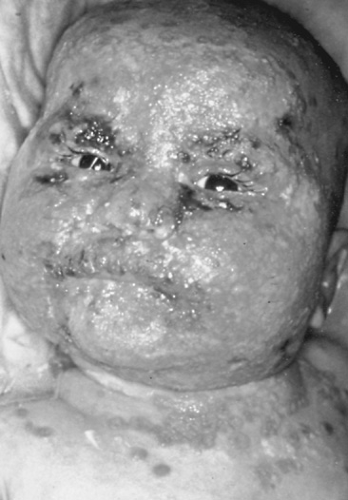 FIGURE 196.1. Extensive herpes simplex virus infection in an infant with atopic eczema (Kaposi varicelliform eruption). (Reproduced with permission from Kohl S. Postnatal herpes simplex virus infection. In: Feigin RD, Cherry JD, eds. Textbook of pediatric infectious diseases, 4th ed. Philadelphia: Saunders, 1998:1709.) |
The epidemiology of HSV is dominated by symptomatic and asymptomatic infection in a huge pool of latently infected individuals. Symptomatic recurrences and asymptomatic shedding ensure the continued spread of HSV. Approximately 1% of individuals shed HSV orally, and 0.2% to 0.5% of women shed HSV genitally at any time. HSV-2 seropositive men and women shed HSV in the genital tract approximately 1% to 5% of the time. The numbers are higher for individuals who are high-risk or immunocompromised. Seroepidemiologic studies reveal that HSV infections are found in all populations. No definite seasonal pattern in HSV infections exists.
Most neonatal HSV infections are acquired from maternal genital strains and, thus, usually are caused by HSV-2. After the neonatal period, HSV-1 infections predominate and, depending on social and economic factors, 40% to 60% of young children of lower socioeconomic status are seropositive by age 5 years. Most such individuals exhibit HSV-1 antibodies by the time they reach adulthood. In one study of adolescents, 62% were seropositive for HSV-1 and 12% for HSV-2.
Studies have documented the acquisition of HSV-1 in child-care nursery or school settings, with clusters of infection and, in some cases, outbreaks of symptomatic illness occurring in as many as 13 children per outbreak. Typically, illness occurs in children 1 to 2 years old, with herpetic gingivostomatitis being the major manifestation. Studies of higher socioeconomic populations reveal seroepidemiologic evidence for HSV-1 infection in only 30% of university students. Reflecting its association with sexual activity, the prevalence of HSV-2 increases at approximately the time of puberty and early adolescence. The percentage of HSV-2-seropositive adults ranges from 20% to 35%, with a 30% increase occurring in the last decade in the United States and a fourfold increase among adolescents.
The incidence of HSV genital infection has increased markedly since the late 1970s. Approximately 1 million new cases occur annually in the United States. In studies of sexually active university students, 4 to 16 per 1,000 acquire genital HSV infection annually. In family health clinics, the rate can be as high as 55 per 1,000.
Risk factors for the acquisition of HSV-2 in North America include gender (female greater than male), race (higher in blacks), lower socioeconomic status, multiple sex partners, failure to use condoms, and bacterial vaginosis. Transmission of HSV-2 from an infected individual to an HSV-2-seronegative individual occurs annually in approximately 10% of stable heterosexual couples. Higher rates occur in transmission from men to women (19%) and to HSV-1- and HSV-2-seronegative women (32%).
The reactivation of latent HSV infection is associated with a variety of influences, including exposure to sunlight (ultraviolet), certain febrile illnesses, local trauma, menstruation, and immunosuppression. These influences, therefore, define additional epidemiologic factors pertinent to HSV infections.
PATHOGENESIS
HSV tends to infect cells of ectodermal origin and, in most cases, initial viral replication occurs at the portal of entry, usually in skin or mucous membranes. The nuclei of infected cells manifest eosinophilic intranuclear inclusions. Because HSV has a predilection for cells that originate in embryonic ectoderm, these viruses may involve the central nervous system (CNS).
The incubation period for primary HSV infection varies from 2 to 20 days in most cases. After primary infection has occurred, the virus remains latent in sensory neural ganglia that innervate portions of the skin or mucous membranes originally involved. Thus, an individual with recurrent HSV almost always experiences reactivation of the HSV lesions in the identical region. In immunologically intact individuals, the recurrence generally is less severe than is the primary infection. In individuals previously infected with one type of virus (e.g., HSV-1, orally), infection with a second type (e.g., HSV-2, genitally) is not prevented but is less severe than in a host who has never been infected with either. Less commonly, an individual can acquire a reinfection with the same type (e.g., a second infection with a new strain of HSV-2 genitally in a patient with preexisting genital HSV-2 infection). Generally, the reinfection is mild and often is dismissed as an endogenous recurrence. These strains can be differentiated by DNA endonuclease restriction analysis of viral isolates. The pathophysiology of recurrent HSV is described in Box 196.2.
CLINICAL MANIFESTATIONS AND COMPLICATIONS
Most infections do not cause significant or specific symptoms. Although they harbor latent HSV, most seropositive persons are unaware of having ever encountered these viruses. The spectrum of symptomatic HSV infections ranges from minor localized recurrences, usually at mucocutaneous junctions, to severe and even fatal illnesses.
Gingivostomatitis
Gingivostomatitis is the most common form of HSV-induced primary illness seen in children. Symptomatic illness may occur
in 30% or more of seropositive infants. Usually, it is seen in young children between ages 6 months and 3 years. In children younger than 6 months old, the presence of residual maternal antibody probably modifies or prevents the appearance of recognizable symptoms in association with HSV infection. Primary gingivostomatitis in children often is acquired from a family member with active primary or recurrent oral HSV infection. Although acute gingivostomatitis caused by HSV occurs relatively infrequently, it still is common enough that most pediatricians should become familiar with the condition and learn to distinguish this infection from herpangina.
in 30% or more of seropositive infants. Usually, it is seen in young children between ages 6 months and 3 years. In children younger than 6 months old, the presence of residual maternal antibody probably modifies or prevents the appearance of recognizable symptoms in association with HSV infection. Primary gingivostomatitis in children often is acquired from a family member with active primary or recurrent oral HSV infection. Although acute gingivostomatitis caused by HSV occurs relatively infrequently, it still is common enough that most pediatricians should become familiar with the condition and learn to distinguish this infection from herpangina.
BOX 196.2. Pathophysiology of Recurrent Herpes Simplex Virus (HSV) Infection
After developing primary HSV infection, immunocompetent individuals have an early nonspecific response followed by a specific immunologic response. The former consists of mobilization of polymorphonuclear and mononuclear leukocytes to the site of infection, release of interferons and other cytokines, and activation of macrophages and natural killer cells. After several days, many types of specific antiviral antibodies are produced. In the second to third weeks of infection, specific cellular immunity manifested by blastogenesis of lymphocytes, production of immune lymphokines (as interferon-gamma, interleukin-2, migration inhibitory factor), a positive delayed hypersensitivity skin test, and T-cell cytotoxicity can be detected. In individuals with cellular immunologic defects (neonates, severely malnourished infants, patients with Wiskott-Aldrich syndrome and other primary immunodeficiencies, and patients receiving transplants or immunosuppressive chemotherapy), primary HSV infection can be a disseminated, life-threatening syndrome, probably because of a defect in cell-mediated immunity of the nonspecific or specific variety.
The immune response to recurrent infection is not well-characterized. It does not appear to be associated with marked alterations in the production of antibody, although fourfold elevations in titer and reemergence of IgA and IgM antiviral antibody may occur. The activity of natural killer cells and production of lymphokines increase, and relative defects in these and the blastogenesis of lymphocytes may be associated with frequent or severe recurrent infection. In the host with cellular defects, recurrences are common events and result in long duration and increased severity but usually do not cause widespread dissemination.
The incubation period covers a few days, and the illness is ushered in by fretful behavior and fever. Usually, affected infants refuse to eat and may even refuse fluids. Vesicular lesions appear on and around the lips, along the gingiva, on the anterior tongue, and on the anterior (hard) palate (Fig. 196.2). Vesicles break down rapidly, and usually lesions appear as 1- to 3-mm shallow gray ulcers on an erythematous base. Generally, the gums are mildly hypertrophic, ulcerated, and erythematous. They may appear friable and frequently bleed on contact. Not uncommonly, vesicles extend about the lips and chin or down the neck in immunologically normal children. Often, the breath emits a foul odor (fetor oris). Affected children experience extreme discomfort and cannot or will not eat; if fluids are refused as well, such children may require hospitalization to maintain adequate hydration. The risk of dehydration occurring is compounded by the fever that usually accompanies this syndrome. The lesions bleed easily and may become covered with a black crust. Often, cervical and submental nodes are swollen and tender. The process evolves for 4 to 5 days, and resolution requires at least an additional week. Autoinoculation may cause lesions on the hands (whitlow) and, less commonly, on the trunk or genital area.
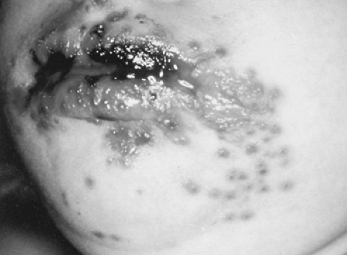 FIGURE 196.2. Primary herpes gingivostomatitis at the ulcerative vesicular stage in a normal toddler. (Reproduced with permission from Kohl S. Postnatal herpes simplex virus infection. In: Feigin RD, Cherry JD, eds. Textbook of pediatric infectious diseases, 4th ed. Philadelphia: Saunders, 1998:1706.) |
HSV gingivostomatitis is differentiated from herpangina, a manifestation of enteroviral infection, by the predominance of ulcers in the anterior and posterior portions of the oropharynx; usually, herpangina is a posterior pharyngeal ulcerative condition. In addition, unlike HSV infection, herpangina often has a more acute onset, shorter duration, and seasonal occurrence. Although enterovirus-mediated hand-foot-and-mouth disease can present with oral ulcers and a vesicular eruption on the distal portion of extremities, its bilaterally symmetric distribution should differentiate it from HSV gingivostomatitis and concurrent HSV autoinoculation of a digit. Severe Stevens-Johnson syndrome (erythema multiforme) may mimic HSV, but the generalized macular rash with bull’s-eye lesions is characteristic of erythema multiforme. HSV can be associated with erythema multiforme (see section, Erythema Multiforme and Herpes Simplex Virus Infection).
In adolescents and especially in college-aged patients, primary HSV infection often manifests as a posterior, occasionally exudative pharyngitis. The characteristic findings are shallow tonsillar ulcers with a gray exudate. In this setting, it must be differentiated from streptococcal infection, Epstein-Barr virus, adenovirus, Arcanobacterium and, rarely, diphtheria- or tularemia-induced pharyngitis. In one study of college students of high socioeconomic status, HSV was diagnosed most often (24%) as the etiology of acute pharyngitis. This manifestation usually is caused by HSV-1, but with the increased frequency of oral-genital sexual practices among both heterosexual and homosexual individuals, HSV-2 pharyngitis is encountered more commonly.
Considering the widespread publicity of HSV as a sexually transmitted disease, health care workers are advised to anticipate patient anxieties when making the diagnosis of HSV oral infection. Unless sexual contact or abuse is suspected, physicians should explain the normal mode of acquisition of oral HSV in young children.
Genital Infections
Primary herpetic vulvovaginitis may occur rarely in very young infants and children if HSV is introduced inadvertently in
handling the genital area with contaminated hands. Moreover, genital herpes may reflect sexual abuse of young children. The occurrence of genital HSV in young children warrants a sensitive and careful appraisal of the family dynamics.
handling the genital area with contaminated hands. Moreover, genital herpes may reflect sexual abuse of young children. The occurrence of genital HSV in young children warrants a sensitive and careful appraisal of the family dynamics.
The incidence of genital infection in adolescents and young adults has increased markedly since the late 1970s; few data address the incidence in children. Approximately 35% to 50% of patients with the first episode of genital herpes report a history of genital HSV in their contacts. HSV-1 accounts for approximately 25% of primary genital HSV infections and 15% to 20% of genital isolates. The incubation period is 2 to 14 days. Primary illness is accompanied by fever, headache, malaise, and myalgias. Other systemic symptoms include an aseptic meningitis syndrome (11% to 35%). Although HSV-2 occasionally may be grown from the CSF, aseptic meningitis syndrome differs from HSV-1 encephalitis in that generally it is mild, self-limited, and not associated with neurologic residua. Local genital symptoms include severe pain, itching, dysuria, vaginal or urethral discharge, and tender inguinal adenopathy. In primary illness, lesions begin as painful vesicles or pustules and progress to wet ulcers and then to healing ulcers with or without crusts. Usually, crusts occur only on squamous epithelium. Lesions tend to last for 2 to 3 weeks before complete healing occurs. Virus shedding occurs for a mean of 11.5 days.
In addition to aseptic meningitis syndrome, the complications of primary HSV genital infection include sacral autonomic nervous dysfunction (manifested as poor rectal sphincter tone, constipation, sacral anesthesia, urinary retention, impotence), extragenital lesions, secondary yeast infections in women, and pharyngitis.
Beyond discomfort and embarrassment, the importance of HSV in the female genital tract relates to the potential impact of the virus on offspring, especially when a child is born to a mother with active viral shedding, particularly in connection with a symptomatic or asymptomatic primary or first episode of maternal infection (see Chapter 78, Herpes Simplex Virus). The presence of genital ulcer lesions, including those caused by HSV infection, increases the risk for the acquisition of human immunodeficiency virus (HIV) infection. An additional consideration is the effect of HSV on the self-image of the young, sexually active patient. Although some individuals cope easily with the illness and the likelihood of having recurrent disease, a sizable number exhibit profound depression, poor self-esteem, complete abstention from sexual activity, and general withdrawal. Self-help groups of individuals who have genital HSV are useful and are located in many cities of the United States.
Other Primary Herpes Simplex Virus Skin Infections
Virtually any part of the skin and mucous membranes may be involved in HSV infections. Often, altered skin provides a portal of entry for HSV. Vesicular lesions spread throughout the affected skin, usually crusting and resolving in approximately 1 week. In normal wrestlers, herpes gladiatorum usually involves the head (73%), extremities (42%), and trunk (28%). The illness accompanying eczema herpeticum can be severe and even fatal, although in most cases, the infection resolves without the administration of specific therapy and leaves no sequelae (see Fig. 196.1). Similar widespread herpetic lesions may occur in skin altered by abrasions or by thermal or chemical burns. In this situation, a secondary fever may occur, usually 1 week to several weeks after the initial insult. Careful inspection of the site or adjacent normal tissue may reveal vesicles or nonspecific ulcerative lesions. Several affected patients who did not receive therapy have died of disseminated HSV infection.
Herpetic whitlow is a painful, erythematous, swollen lesion occurring at a site of broken skin on the terminal phalanx of fingers (69%) and thumb (21%). Less commonly, toes are involved. The painful white swellings appear to be filled with pus, but, when opened for drainage, they are found to contain little fluid and no purulent material. Occasionally, the whitlow, which may persist for 7 to 10 days, initially is accompanied by a few vesicles that may give a clue to the etiology of the infection. Whitlows are seen in four typical situations. First, infants with herpetic gingivostomatitis may autoinoculate their fingers (Fig. 196.3). Second, whitlows are encountered in infants without obvious oral disease, sometimes caused by infected adults kissing their children’s fingers. Third, in sexually active patients, more often the whitlow is a manifestation of concurrent genital disease, which should be investigated through appropriate history and physical examination. Fourth, dentists, respiratory therapists, nurses, and pediatricians who sometimes examine oral cavities or handle secretion-contaminated material without wearing gloves are at risk for developing herpetic whitlows. Because of the epidemiology of HSV, usually whitlows in children are caused by HSV-1. Importantly, the herpetic condition should be diagnosed because usually it is confused with a bacterial felon or paronychia and is incised and drained. This outcome is not indicated in the therapy of HSV whitlow. Only a needle aspiration and culture are necessary for diagnosis of herpetic whitlow. Appropriate infection-control measures will reduce the spread of virus due to whitlows.
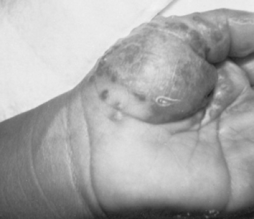 FIGURE 196.3. Herpetic whitlow in a toddler with oral herpes simplex virus infection. (Reproduced with permission from Kohl S. Postnatal herpes simplex virus infection. In: Feigin RD, Cherry JD, eds. Textbook of pediatric infectious diseases, 4th ed. Philadelphia: Saunders, 1998:1710.) |
Herpes Simplex Virus of the Eye
Primary HSV infection of the eye may manifest as a blepharitis or a follicular conjunctivitis, often accompanied by preauricular lymphadenopathy. If restricted to the conjunctiva, the infection, which can be accompanied by vesicular herpetic lesions elsewhere on the face or in the nose or mouth, usually resolves without sequelae. Herpetic infection of the eye may, however, progress to involve the cornea, with more serious potential consequences. For this reason, ophthalmologists always should examine and evaluate such cases.
Corneal involvement by HSV may manifest initially with minute vesicles at the corneal margin. The progress of corneal infection (best seen with the use of topical fluorescein dye) is
marked by the appearance of branching lesions (a dendritic pattern) or the less diagnostic irregular (ameboid or geographic) ulcer. The affected child complains of severe photophobia, blurred vision, chemosis, and lacrimation. Primary eye infection may include stromal involvement, uveitis, and (rarely) retinitis. Spontaneous healing, which generally requires 2 to 3 weeks, can be expedited by the use of antiviral therapy (see section, Therapy, page 1256). Corticosteroids are contraindicated. The risk of developing visual impairment is enhanced with recurrences. With each bout of infection, the dendritic ulcers are more extensive and more liable to result in scarring and loss of sight. Rarely, in the immunocompromised host, HSV has been associated with retinal necrosis.
marked by the appearance of branching lesions (a dendritic pattern) or the less diagnostic irregular (ameboid or geographic) ulcer. The affected child complains of severe photophobia, blurred vision, chemosis, and lacrimation. Primary eye infection may include stromal involvement, uveitis, and (rarely) retinitis. Spontaneous healing, which generally requires 2 to 3 weeks, can be expedited by the use of antiviral therapy (see section, Therapy, page 1256). Corticosteroids are contraindicated. The risk of developing visual impairment is enhanced with recurrences. With each bout of infection, the dendritic ulcers are more extensive and more liable to result in scarring and loss of sight. Rarely, in the immunocompromised host, HSV has been associated with retinal necrosis.
Herpes Simplex Virus Infections of the Central Nervous System
HSV is the most common identifiable cause of sporadic encephalitis, which usually is very serious. It accounts for 2% to 5% of all cases of encephalitis in the United States and for as many as 20% of all etiologic diagnoses (60% to 70% of cases of encephalitis remaining without a diagnosis). The case-fatality rate associated with untreated HSV encephalitis is approximately 70%, and survivors generally exhibit considerable permanent neurologic disability. The spread of HSV-1 to the CNS seems to proceed via neurogenic pathways. Although HSV encephalitis may involve virtually any area of the brain, it shows a striking tendency to involve the frontal and temporal lobes after the neonatal period.
An important step is to differentiate the HSV-induced aseptic meningitis syndrome—usually caused by HSV-2 and usually a complication of primary genital infection—from HSV encephalitis. In the former, signs of meningitis, including headache, photophobia, and stiff neck, appear shortly after genital lesions are noted. Usually, seizures and focal CNS findings are absent. An examination of the cerebrospinal fluid (CSF) reveals a lymphocytosis, with 300 to 2,600 white blood cells (WBCs) per cubic millimeter and sometimes a low glucose level. This syndrome may recur with genital recurrences. Usually, complete recovery occurs without the administration of specific therapy. HSV occasionally may be grown from the CSF.
In contrast to meningitis, HSV encephalitis is a highly lethal disease. In 96% of cases, it is caused by HSV-1. It may be a result of primary (30%) or recurrent (70%) infection. A larger percentage of cases of HSV encephalitis in younger individuals probably is due to primary infection. One-third of cases occur in the pediatric age range. As in most manifestations of HSV infection, but unlike most other common forms of viral encephalitis (enterovirus, arbovirus), HSV encephalitis has no seasonality. It is an acute illness with fever, malaise, irritability, and nonspecific symptoms lasting 1 to 7 days, progressing to signs and symptoms of focal CNS involvement in 3 to 7 days, and finally to coma and death (Table 196.1). The advent of sensitive HSV DNA polymerase chain reaction (PCR) diagnosis has confirmed that most cases are focal, although mild and atypical presentations occur more commonly than was thought. Fever and altered behavior in any child should evoke suspicion of encephalitis. Meningeal signs are uncommon findings. No correlation exists between the isolation of HSV from sites extrinsic to the CNS (e.g., the oropharynx or genital tract) and the diagnosis of HSV encephalitis. Thus, the presence of oral or genital lesions is not helpful in the diagnosis or exclusion of HSV encephalitis. Both identical and discordant viruses have been isolated simultaneously from the brain and oral secretions.
The CSF generally reveals a pleocytosis with as many as 2,000 WBCs per cubic millimeter, usually (80% of cases) more than 50 WBCs per cubic millimeter. In 90% of cases, more than 60% of cells are lymphocytes. Early in the infection, neutrophils may predominate. In 75% to 85% of cases, red blood cells, reflecting the hemorrhagic necrosis, are seen in the CSF. Between 5% and 25% of patients have hypoglycorrhachia, and 80% have elevated CSF protein levels (median, 80 mg/dL), which rise to striking levels with progression of the disease. The CSF is normal in 2% to 3% of patients with early HSV encephalitis. HSV almost never is grown from lumbar CSF and rarely from ventricular fluid. Thus, whereas the CSF examination is helpful, it is not diagnostic of HSV encephalitis unless PCR is used to detect HSV DNA.
TABLE 196.1. HISTORICAL AND CLINICAL FINDINGS IN HERPES SIMPLEX VIRUS ENCEPHALITIS | ||||||||||||||||||||||||||||||||||||||||||||
|---|---|---|---|---|---|---|---|---|---|---|---|---|---|---|---|---|---|---|---|---|---|---|---|---|---|---|---|---|---|---|---|---|---|---|---|---|---|---|---|---|---|---|---|---|
| ||||||||||||||||||||||||||||||||||||||||||||
The usefulness of neurodiagnostic tests is limited. The electroencephalography (EEG) probably is one of the more useful tests. A “typical” pattern of unilateral or bilateral (poor prognosis) periodic focal spikes against a background of slow (flattened) activity (paroxysmal lateral epileptiform discharges) is associated with HSV encephalitis. Other findings include large-amplitude irregular slow activity, sharp waves, and variable spikes. In 80% to 90% of patients, the EEG finding is not only abnormal but localizing. This test is one of the earliest localizing laboratory studies. The brain scan or computed tomography (CT) is less helpful early in the illness. Late in the illness, the CT appearance may be characteristic [i.e., low-density, contrast-enhanced lesions in the temporal area, mass effect, edema, and hemorrhage (Fig. 196.4)], but early in the illness, when diagnosis is critical, often the CT appearance is unremarkable. An abnormal CT scan is a poor prognostic factor. Magnetic resonance imaging (MRI), an early sensitive test for localizing HSV encephalitis (Fig. 196.5), is the radiologic technique of choice for making an early diagnosis of HSV encephalitis. The finding of focal abnormality on EEG, MRI, CT, or radionuclide brain scan is significantly more likely to occur in HSV encephalitis than in those other illnesses with which it is confused.
Related posts:

Stay updated, free articles. Join our Telegram channel

Full access? Get Clinical Tree


