Posteromedial and Posterolateral Release for the Treatment of Resistant Clubfoot
Richard S. Davidson
DEFINITION
Clubfoot, or talipes equinovarus, is a congenital or acquired deformity in which the foot is stiffly positioned in hindfoot equinus and varus and forefoot varus, supination, and plantarflexion.8
When the deformity is not corrected, the patient limps, bearing weight on the lateral forefoot. This can limit ambulation and lead to foot and ankle pain, abnormal calluses, and ulcers and infections.2
The deformity may present as an isolated or syndromic birth defect. Clubfoot has been documented in conjunction with diagnoses of polio, spina bifida, cerebral palsy as well as other disorders.6
ANATOMY
Clubfoot deformity begins as soft tissue imbalance and contractures altering the positions predominantly of the talus, calcaneus, and navicular as well as their corresponding articulations.6
The displacement of these bones differs, producing varying amounts of four different positional deformities: cavus, adductus, varus, and equinus.3
Eventually, the abnormal forces and positions lead to plantarflexion and medialization of the talar neck.6
Weakness and underdevelopment of the foot and calf result to varying degrees.5
Although radiographic measurements are hard to reproduce, the anteroposterior (AP) and lateral talocalcaneal angles are reduced from about 28 degrees to about 5 degrees in children with clubfoot.6
PATHOGENESIS
The exact cause of clubfoot remains unknown. Many theories can be found in the literature. The cause is probably multifactorial and includes some extent of the following2, 5, 6, 7:
Primary germ plasma defect: Initial investigations into multiple cases of clubfoot speculate that the consistent bony deformity is caused by primary bone dysplasia.
Uterine restriction: A reduced amount of amniotic fluid causes limited fetal foot movement and, incidentally, clubfoot.
Bone-joint hypothesis: The cause of the deformity is abnormalities in the ossification of the bones of the foot.
Connective tissue hypothesis: Degeneration in the connective tissues of the skeletally immature foot causes clubfoot.
Vascular hypothesis: Muscle wasting has been documented in most children with idiopathic congenital talipes equinovarus. Type 1 fiber predominance and grouping also coincides with most cases of clubfoot.
Neurologic complication: Clubfoot is seen in conjunction with a long list of neurologic disorders, including spina bifida, anencephaly, hydrocephaly, and so forth.
Developmental arrest hypothesis: Due to a noted similarity between clubfeet and the embryonic foot at the beginning of the second month of fetal development, it has been suggested that the maturation of the fetal foot was arrested while under genetic control.
Genetics: This is the most probable cause, as agreed on by many physicians; a family history of talipes equinovarus has been documented in a majority of the reported cases.
NATURAL HISTORY
The overall incidence of talipes equinovarus is about one per thousand live births but varies with sex and race: It is more commonly seen in boys, and there is a high frequency of affected children in Polynesian cultures.7
Untreated, the deformity leads to limping, abnormal calluses due to weight bearing on the lateral forefoot, atrophy and hypoplasia due to disuse, and pain.
Research has been extensive regarding the appropriate approach to treating clubfoot, but few long-term comparative studies exist. Although in the past century extensive surgery predominated, it is now believed that extensive surgical techniques are necessary in fewer than 5% of cases.
Currently, the most popular treatment of clubfoot follows Ponseti method, which was not generally accepted until his review article of 1992 in which he demonstrated results similar to those of more extensive surgery with fewer complications. The technique has gained such popularity that most pediatric orthopaedists throughout the world are employing Ponseti basic principles of manipulation, casting, and minimal operative treatment of the clubfoot.
PATIENT HISTORY AND PHYSICAL FINDINGS
The deformity of clubfoot is identified at birth as unilateral or bilateral hindfoot equinus and varus and midfoot supination, varus, and equinus.
To perform the examination, the leg is extended at the knee and the foot is then dorsiflexed. The foot-to-tibia angle is measured to assess the amount of equinus in the frontal plane and the amount of heel varus in the sagittal plane.
The dorsolateral aspect of the midfoot is palpated to locate the talar head. The forefoot is then manipulated to determine if the forefoot can be reduced onto the talar head.
The lateral rotation of the foot-thigh angle can be assessed by flexing the knee and ankle to 90 degrees and gently laterally rotating the foot. The angle is measured.
These examinations do not determine a classification but rather the stiffness of the foot and the amount of improvement attained with serial casting and surgical intervention.
The clinician should investigate associated anomalies, such as spina bifida, spasticity, muscular dystrophy, arthrogryposis, and so forth. By understanding the cause, the likelihood of treatment success can be predicted.
The clinician should observe the shape and size of the foot. The clubfoot is generally shorter and wider than a normal foot.
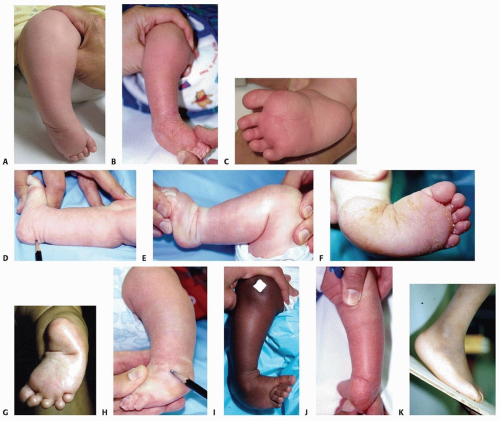
Examination reveals equinus and varus of the ankle and midfoot. Creases or clefts are seen at the midfoot and ankle. Calf atrophy is expected, particularly in the older child (FIG 1A-C).
Treatment may be altered depending on the presentation of the clubfoot.
Range of motion: equinus
Ankle motion (dorsiflexion and plantarflexion) is assessed in both knee extension and flexion. The os calcis may remain in equinus (by palpation) even though the heel pad appears to come out of equinus (by observation) (FIG 1D,E). This is the so-called empty heel pad sign.
Therefore, the foot may “look” as if the equinus is corrected, but the physician must palpate it to know for sure.
Range of motion: subtalar joint
Range of motion is difficult to measure. The resting alignment of the heel to the talus is usually varus in the untreated clubfoot and 5 to 10 degrees of valgus in the corrected foot. The clinician looks at the sole of the foot to observe midfoot varus. The sole is manipulated to see how flexible it is (FIG 1F,G).
Overcorrection of the heel into valgus can lead to painful pronation. Residual varus of the lateral border of the foot may be due to subtalar rotation, varus of the calcaneus, medialization of the cuboid on the calcaneus, or varus deformity of the metatarsals. Correction may be required at the site of deformity.
Range of motion: forefoot on the talar head
The foot is palpated dorsolaterally at the lateral midfoot. It usually is lined up with the patella, although plantarflexed. Manipulation is used to reduce the forefoot (FIG 1H).
The more difficult it is to reduce the forefoot onto the talar head, the stiffer the deformity.
Forefoot supination
The clinician observes that the forefoot of the clubfoot appears supinated with respect to the tibia. However, supination relates to the position of the forefoot to the hindfoot (FIG 1I,J).
If the forefoot appears 30 degrees supinated to the tibia and there is 30 degrees varus to the hindfoot varus, then
the deformity is hindfoot varus and not supination, that is, the forefoot is properly aligned to the hindfoot and there is no supination.
It is important to know where this deformity is. Errors in this assessment may lead the surgeon to overcorrect the midfoot or surgically create a pronation deformity.
Forefoot plantarflexion
The physician begins with palpation of the medial column from the first metatarsal to the talar head. Plantarflexion of the forefoot on the hindfoot is measured.
In the operated foot, the physician checks for dorsolateral subluxation or dislocation of the navicular on the talar head (FIG 1K).
Deformity must be corrected where it is. This assessment, in conjunction with radiographs, will help to assess its location in the soft tissues, the ankle, the bone, or the joints (such as subluxation of the talonavicular joint).
IMAGING AND OTHER DIAGNOSTIC STUDIES
Sonograms may be used to diagnose clubfoot prenatally.5 Although no prenatal treatment is available, many parents want to know the diagnosis of clubfoot so they can learn about the natural history and treatment options available to them.
The prenatal ultrasonographic diagnosis of clubfoot may be made if the bones of the lower leg are in the same plane as the plantar surface of the fetal foot. To ensure a correct diagnosis, images in which the leg is extended away from the wall of the uterus should be obtained.5
This deformity may be seen as early as 12 or 13 weeks.5
Stay updated, free articles. Join our Telegram channel

Full access? Get Clinical Tree


