Surgical Management of Blount Disease
Richard S. Davidson
DEFINITION
Blount disease, also known as idiopathic tibia vara and osteochondritis deformans tibiae, is characterized by abnormal growth of the proximal tibia physis with progressive varus deformity.
Blount disease is classified into three types based on age of clinical onset: infantile (0 to 3 years), juvenile (4 to 10 years), and adolescent (11 years and older).9
Infantile tibia vara is most prevalent in African American females and is associated with obesity, internal tibial torsion, and leg length discrepancy. Radiographs reveal a prominent medial metaphyseal beak, and the origin of the varus deformity is in the proximal tibia only. About 80% of cases are bilateral, and the potential for deformity is the greatest in this group.
Adolescent tibia vara is most prevalent in African American males with marked obesity, minimal internal tibial torsion, mild medial collateral ligament laxity, and mild leg length discrepancy. The site of the deformity is in the proximal tibia and sometimes in the distal femur as well. About 50% of cases are bilateral, and pain rather than deformity is more commonly the presenting complaint.
ANATOMY7
When evaluating patients with Blount disease, the normal development of the tibiofemoral angle in children must be considered.
The normal tibiofemoral angle in newborns is approximately 15 degrees varus. It decreases with growth, so that the tibiofemoral angle approaches 0 degrees around 18 months of age.
The tibiofemoral angle progresses to maximum valgus around 3 years of age and then decreases until adult physiologic valgus is achieved between 7 years of age and skeletal maturity.
One standard deviation of the anatomic tibiofemoral angle throughout growth is approximately 8 degrees.
PATHOGENESIS7
Blount disease is likely due to a combination of genetic factors and a cycle of increased stress across the medial physis, which leads to decreased medial endochondral ossification, further varus deformity, and, subsequently, further medial physeal stress. The medial physeal stress is aggravated by obesity and progressive genu varum.
Histopathologic studies of infantile and late-onset tibia vara are similar to those of patients with slipped capital femoral epiphysis. Findings include fissuring and clefts in the physis, fibrovascular and cartilaginous repair at the physeal-metaphyseal junction, foci of necrotic cartilage, and marked disorganization of the medial degenerative physeal zone.
These findings are consistent with an arrest of the normal endochondral growth mechanism.
NATURAL HISTORY
A varus alignment of the lower extremity places excess stress on the medial compartment of the knee. This stress places the knee at increased risk for arthritis.
The goal of intervention is to restore the normal anatomic orientation of the knee and ankle joints and to restore the normal mechanical axis of the leg.
PATIENT HISTORY AND PHYSICAL FINDINGS
The chief complaint in infantile tibia vara is usually deformity. In late-onset tibia vara, in contrast, knee pain is the primary complaint. The characteristics of the pain should be elicited.
Patients may exhibit a limp, with or without a leg length discrepancy. Observe the patient’s gaiting, noting a limp or lateral thrust.
The mechanical axis of the lower leg is in varus. Genu recurvatum and internal tibial torsion may be present as well.
Inspect the sagittal profile for the presence of genu recurvatum; if present, it may be necessary to address it at the time of surgery.
The Q angle provides a clinical estimate of the anatomic tibiofemoral angle.
Range of motion and collateral ligament laxity also should be assessed.
IMAGING AND OTHER DIAGNOSTIC STUDIES
Anteroposterior (AP) long-leg radiographs (which include the hips, knees, and ankles) should be obtained (FIG 1). The patella (not the foot) must be pointing forward.
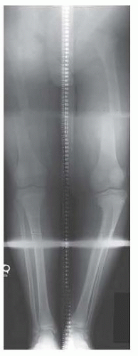
FIG 1 • Orthoradiograph of patient with adolescent Blount disease.
Infantile Blount disease has several characteristic radiographic findings.
To help differentiate infantile Blount disease from physiologic varus, the metaphyseal-diaphyseal angle is drawn. A metaphyseal-diaphyseal angle less than 10 degrees is consistent with physiologic varus, whereas an angle of more than 16 degrees is consistent with infantile Blount disease.
Acute angulation of the medial proximal tibia, medial beaking, fragmentation of the medial metaphysis, progressive varus, and unilateral involvement are consistent with infantile Blount disease.
Care must be taken that the radiographs are taken with the patella forward.
If tibial torsion is present, the feet must cross medially so that the patella is forward. The medial and lateral flares of the distal femurs will be equal if the patella is forward.
The proximal tibia is examined to determine the Langenskiöld stage.
Stage I: age younger than 3 years; medial and distal beaking of metaphysis with irregularity of entire metaphysis
Stage II: age 2.5 to 4 years; sharp anteromedial depression in ossification line of wedge-shaped medial metaphysis
Stage III: age 4 to 6 years; deepening of metaphyseal beak
Stage IV: age 5 to 10 years; enlargement of epiphysis
Stage V: age 9 to 11 years; cleft in epiphysis, appearance of double epiphysis
Stage VI: age 10 to 13 years; closure of medial proximal tibial physis
Late-onset Blount disease is characterized by less obvious changes in the proximal tibia.
These changes include wedging of the medial portion of the epiphysis, a mild posteromedial articular depression, a serpiginous curved physis of variable width, and mild or no fragmentation of the proximal medial metaphysis.
Radiographic analysis for deformity has been well described by Paley et al.4
The magnitude of the overall lower extremity malalignment can be determined by the anatomic tibiofemoral angle or the mechanical axis deviation. The anatomic tibiofemoral angle is the angle between the midshaft lines of the femur and the tibia. The mechanical axis deviation is the distance from the center of the knee to the mechanical axis line of the leg.
Analysis of the frontal plane deformity begins with the malalignment test.
The mechanical axis line is drawn from the center of the hip to the midpoint of the ankle plafond.
To identify whether the source of the deformity is the femur, the tibia, or both, joint orientation angles are measured.
The mechanical lateral distal femoral angle (mLDFA, normal value is 85 to 90 degrees) and medial proximal tibial angles (MPTA, normal value is 85 to 90 degrees) are measured to determine which is/are abnormal.
The joint line convergence angle is measured to determine whether the joint line is an additional source of deformity.
If the midpoints of the femur and tibia are over 3 mm apart, then frontal plane subluxation is a source of deformity as well.
Finally, the joint lines are inspected for intra-articular sources of deformity.
The malorientation test is applied to the ankle and hip to determine whether these joints are oriented normally to the mechanical axis line.
Abnormal joint orientation angles indicate which joints are contributing to the deformity.
Sagittal plane radiographs are obtained and analyzed as appropriate.
Leg lengths are measured in order to identify a leg length discrepancy.
The location of the deformity point, or center of rotation of angulation (CORA), is identified during preoperative planning.
DIFFERENTIAL DIAGNOSIS
Physiologic varus
Pathologic causes
Rickets
Skeletal dysplasias
Focal fibrocartilaginous dysplasia
Renal osteodystrophy
Osteogenesis imperfecta
NONOPERATIVE MANAGEMENT
Nonoperative treatment with bracing may be indicated in patients with infantile Blount disease.
Bracing should be considered for varus deformity greater than 15 degrees in children older than 2 years of age with Langenskiöld stage I or II Blount disease.2
Bracing usually is not helpful in obese African American girls older than the age of 3 years.
Nonoperative treatment with bracing is not successful in adolescent Blount disease.
SURGICAL MANAGEMENT
The surgical treatment of infantile Blount disease is distinct from that for adolescent Blount disease.
In patients with infantile Blount disease, the proximal tibial physis has several years of growth remaining. A proximal tibial osteotomy should be performed with the goal of correcting the anatomic tibiofemoral angle to within 5 degrees of neutral. In addition to the osteotomy, medial proximal tibial physeal bar resection, lateral proximal tibial hemiepiphysiodesis (guided growth plates), or tibial plateau elevation can be performed to improve the alignment of the physis and to allow for proper future growth.
Definitive surgery for infantile Blount disease should be done before 5 years of age because recurrence may develop if surgery is performed after this age.
In patients with adolescent Blount disease, treatment options are hemiepiphysiodesis and osteotomy. However, if insufficient growth remains for hemiepiphysiodesis to be effective, osteotomy is the best option for correction of the deformity. Hemiepiphysiodesis of an already short limb may leave the patient with a significant limb length inequality. If such limb length inequality will require
osteotomy for lengthening, the tibia vara should be corrected by osteotomy for angular and linear correction with external fixation.
The objective of the osteotomy is to obtain a neutral mechanical axis with a horizontal knee joint. Many different types of osteotomies have been described for the treatment of adolescent Blount disease, including opening and closing wedge osteotomies, dome osteotomies, and oblique osteotomies.
Following the osteotomy, fixation may be achieved with external or internal fixation. The use of cast immobilization alone has been associated with a loss of correction.
Internal fixation after osteotomy for Blount disease has been associated with problems. Loder et al3 reported poor results in patients treated with internal fixation and noted many were internally fixed in malposition, likely due to difficulty in assessing intraoperative alignment. Crossed K-wires have been associated with a loss of fixation. The use of plates has been associated with stress shielding, delayed and nonunion, and hardware breakage, and requires a second surgical procedure to remove the implant.
External fixation allows for acute or gradual correction and for later adjustments as clinically and radiographically indicated. In addition, external fixation allows for correction of the coexistent leg length discrepancy. Price et al5 reported the successful use of dynamic external fixation to stabilize osteotomies for tibia vara without supplemental casting. Monolateral, hybrid, or circular external fixators may be used.Related posts:

Stay updated, free articles. Join our Telegram channel

Full access? Get Clinical Tree


