Posterolateral Corner and Fibular Collateral Ligament Reconstruction
E. Lyle Cain Jr.
William G. Clancy Jr.
Posterolateral rotatory instability (PLRI), unlike anterior cruciate ligament (ACL) deficiency, is a relatively infrequently encountered instability pattern that has historically been underdiagnosed and poorly treated. Because this instability pattern is uncommon, it is frequently not diagnosed at the time of initial injury, often presenting as chronic disability. Posterolateral corner injury may result in the worst functional instability pattern of any type of knee ligament injury. These injuries produce excessive tibial external rotation, posterior tibial translation, and varus laxity. Excessive external rotation of the tibia allows for an unwinding moment on the cruciate ligaments, which enhances joint laxity and adversely affects the cruciate ligament reconstruction or the native ligament mechanics. External tibial rotation brings the ACL tibial insertion laterally toward its lateral femoral condyle origin, producing laxity in the ACL. Similarly, external tibial rotation brings the posterior cruciate ligament (PCL) tibial insertion medially toward the medial femoral condyle origin, producing laxity of the PCL.
INDICATIONS/CONTRAINDICATIONS
Posterolateral rotatory instability of the knee, defined has excessive external tibial rotation producing functional instability, can be one of the most disabling of all knee instability patterns. The posterolateral corner consists anatomically of one dynamic stabilizer and several static stabilizers that contribute to a functional stability of the knee. The popliteus muscle tendon complex is the primary dynamic stabilizer, and the popliteal fibular ligament has received added attention recently in the orthopaedic literature as a static stabilizer. The other static structures important for stability include the fibular collateral ligament (FCL) complex as well as the arcuate ligament, posterolateral capsule, and PCL. The importance of PLRI is that if not diagnosed and treated properly, it may lead to failure of other ligament reconstructions, particularly ACL or PCL reconstruction procedures. Harner and colleagues have found that sectioning of the posterolateral structures of the knee resulted in a 150% increase in PCL forces in cadaveric knees. Posterolateral rotatory instability can be classified into one of five types according to the etiology and associated instability patterns with or without varus laxity or thrust.
The first type of PLRI is traumatic. This often occurs secondary to a significant contact injury, most commonly a varus blow from the medial surface of the knee causing a lateral or posterolateral corner tear. The lateral stabilizing instructions of the knee are frequently torn concomitantly, including the fibular collateral ligament as well as other ligamentous structures including the ACL, PCL, or both. With traumatic PLRI, treatment of the other ligament injuries such as cruciate ligament injury will correct anterior and posterior translation of the knee; however, rotational instability may persist. Failure to address PLRI may ultimately lead to failure of ACL or PCL reconstruction. The second type PLRI is known as symptomatic physiologic instability. This type of PLRI is most commonly caused by relatively minor injury occurring in persons already having symmetric excessive external rotation in both knees. However, it may occur without history of even minor trauma, similar to the development of multidirectional instability of the shoulder. We believe that this type of PLRI may be due to electromechanical dissociation of the popliteus muscle, similar to that described by Rowe in the multidirectionally unstable shoulder. In this typem of PLRI, the cruciate ligaments are normal as is the fibular collateral ligament. Lateral (varus) laxity is similar to the ininvolved leg. This rotational instability produces clinical symptoms and history of giving way and instability, particularly with rotational maneuvers, because of excessive lateral tibial rotation with pivoting maneuvers. The diagnosis of symptomatic physiologic instability is often overlooked because of intact cruciate ligaments and a relatively minor trauma necessary to produce this clinical instability. We have observed physiologic asymptomatic excessive external tibial rotation in approximately 10% to 15% of normal knees. Cooper and others have reported excessive physiologic external tibial rotation in 35% of patients. It is important to note that this type of PLRI is typically bilateral and usually does not occur with any significant varus laxity.
The third type of PLRI is combined traumatic cruciate injury with physiologic posterolateral laxity. This occurs with isolated tearing of either the ACL or PCL with concomitant low-grade injury to the posterolateral corner in an individual already having excessive physiologic external rotation. We assume this injury to be the case whenever there is a 3+ posterior drawer present along with excessive tibial external rotation equal to the contralateral side with a PCL injury. Clinically, there is often no increased varus laxity nor is there any injury visible to the popliteus tendon or fibular collateral ligament seen on magnetic resonance imaging (MRI). With concomitant tearing of the ACL, the ACL deficiency and posterolateral rotatory instability often negate each other. Reconstruction of the ACL without attention to the posterolateral corner eliminates the anterior posterior instability but effectively increases the dysfunction of posterolateral excessive rotation of PLRI. Sometimes this occurs to such extent that the individual complains of symptoms with instability worse than was felt before ACL surgery. It is very difficult to determine whether the physiologic posterolateral laxity is pathologic and whether it should be addressed during ACL reconstruction. After the ACL graft has been fixed during reconstruction, the presence of a mild increase in Lachman or pivot glide strongly suggests that the PLRI is pathologic and should be corrected surgically.
In cases of isolated PCL injury with physiologic external rotation deformity, isolated reconstruction of the PCL without reconstruction of the posterolateral corner results in persistence of external rotation excess and posterior tibial sag. Increased external rotation causes the tibial insertion of the PCL to rotate medially and anteriorly, shortening the distance of insertion of the PCL on the medial femoral condyle, and producing functional laxity of the PCL although the PCL reconstruction may be intact. Therefore, symptoms may persist after PCL reconstruction if PLRI is not addressed.
The fourth type of PLRI includes posterolateral instability with varus alignment and varus thrust. This clinical situation occurs in the patient who has an injury to the posterolateral structures with or without fibular collateral ligament injury in a varus knee. When this patient is weightbearing, they have a significant varus thrust which leads to worsening of the PLRI and varus instability. With this situation, medial compartment degenerative changes and joint space narrowing are often present as well as the native varus alignment. We often perform valgus osteotomy in these patients to correct both the varus position as well as a varus thrust prior to, or in combination with, posterolateral reconstruction.
The fifth type of PLRI is posterolateral instability with medial and lateral laxity. This is a rare entity but is seen when the femur has a medial then lateral shift on the tibia during the stance phase of gait. This could be significant for posterolateral laxity with varus thrust; however, it is a straight medial-lateral instability pattern not associated posterolateral corner injury. Most likely, this represents musculoelectrical disassociation similar to pathology in some patients with multidirectional instability of the shoulder and will not benefit from present surgical techniques to reconstruct the posterolateral corner.
Our current indications for posterolateral cruciate reconstruction or fibular collateral ligament reconstruction include a symptomatic knee with any of the PLRI patterns classified previously. The most common patient in our practice presents with either ACL or PCL injury with combined traumatic posterolateral corner injury as well. Any findings on MRI or examination of ecchymosis or swelling along the posterolateral structures of the knee including popliteus, fibular collateral ligament, or posterolateral capsule should be assumed to have posterolateral rotatory instability until proven otherwise by appropriate examination. In the acute setting, we generally repair all the injured structures including the fibular collateral ligament, biceps femorus muscle tendon complex, iliotibial band, popliteus tendon complex, as well as the posterolateral capsule. In many acute cases, the popliteus muscle tendon complex has been damaged to the extent that is not reasonable to repair this structure. Injuries to the popliteus often occur in the muscle tendon junction and are not amenable to acute repair. In these acute settings with posterolateral corner instability and poor tissue quality or muscle tendon junction popliteus tear, we generally perform an acute posterolateral cruciate reconstruction using allograft Achilles tendon. In the chronic situation, we perform posterolateral cruciate reconstruction with or without fibular collateral ligament reconstruction using Achilles tendon allograft.
Several authors have described techniques for either reattaching, repairing, or reconstructing the posterolateral and fibular collateral structures. Hughston’s posterolateral reconstruction, described as anterior and superior advancement of the lateral gastrocnemius tendon, superior posterolateral capsule, fibular collateral ligament, and popliteus tendon, is possible only with intact native structures and has not been met with much success in our experience. Jakob and associates have recommended recession of the popliteus tendon insertion on the femur, but this often fails to improve static or functional stability because of damage to the structures distal to that insertion site. Until 1996, biceps femoris tendon tenodesis and rerouting at the lateral femoral condyle was the senior author’s (WCG) preference for correction of mild to moderate PLRI without the presence of varus thrust. Although this procedure led to predictable and satisfactory results in the mild and moderate cases, it required an intact biceps muscle tendon complex and often required hardware removal at a second surgical procedure. We have performed the posterolateral cruciate reconstruction in severe cases since 1979, and have employed it exclusively in mild, moderate, or severe PLRI since 1996.
Our current preference for acute augmentation or chronic injuries to the posterolateral corner is the posterolateral cruciate reconstruction (PLCR) with one-half Achilles tendon allograft as well as fibular collateral ligament reconstruction with the opposite half of the Achilles tendon allograft. Posterolateral cruciate reconstruction differs from the Hughston or Jakob reconstructions in that PLCR does not rely on healthy native tissues. Immediate knee motion is possible, and hardware removal is usually unnecessary. However, posterolateral cruciate reconstruction alone does not correct for straight lateral (varus) laxity. Therefore, if varus laxity is additionally present with PLRI, the fibular collateral ligament is reconstructed as well.Valgus tibial osteotomy may be considered in conjunction with PLCR or other cruciate ligament procedures in some cases. These patients typically have varus laxity with medial joint space narrowing and a varus thrust in the stance phase of gait. Failure to correct the varus alignment may cause stretching and eventual failure of the posterolateral structures, as suggested by Noyes. At the time of surgery, the osteotomy should be performed first followed by either concomitant or delayed posterolateral cruciate reconstruction and fibular collateral ligament reconstruction.
PREOPERATIVE PLANNING
A proper preoperative evaluation is essential to correctly evaluate the instability pattern for posterolateral rotatory instability.
History
The typical history of patients with acute PLRI is significant for a discrete injury with a blow to the medial side of the knee resulting in varus force, often combined with hyperextension (Fig. 21-1). The mechanism of injury should alert the examiner to look for disruption of the posterolateral structures, the lateral structures, especially the fibular collateral ligament, and associated ACL or PCL injury.
In the chronically disabled individual, the history of significant remote trauma is frequently elucidated but the mechanism is often unclear. More importantly, the individual expresses a very frequent sense of instability with minimal activities, causing giving way that may be associated with
ACL instability. In addition, each episode of giving way is usually accompanied by pain along the posterolateral corner of the knee.
ACL instability. In addition, each episode of giving way is usually accompanied by pain along the posterolateral corner of the knee.
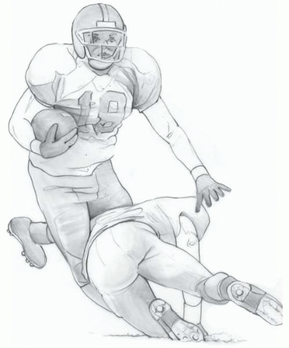 FIGURE 21-1 Typical mechanism for acute posterolateral corner and ACL injury by a blow to the anteromedial tibia causing varus and hyperextension. |
Physical Examination
In the acutely injured knee, findings often include marked rotational and varus instability at 30 degrees of flexion (with fibular collateral and posterolateral injury). If significant varus instability is present at full extension (0 degrees) or 90 degrees flexion, suspicion for disruption of one of the cruciate ligaments must be evaluated. The amount of excessive external rotation can be measured with the pateint supine with hips flexed 90 degrees and the knee flexed 30 degrees. The lower leg is externally rotated and heel and foot position is noted for both legs to determine the amount of relative side to side exernal rotation. The knee is then flexed to 90 degrees and similar external rotation is performed. Some believe that the amount of external rotation is best assessed in the prone position with foot external rotation at 30 and 90 degrees knee flexion. We believe that a more accurate method to measure excessive external rotation is done by placing the patient supine with the hips flexed 90 degrees and knees 30 degrees. The lateral femoral epicondyle and anterior and posterior borders of the fibular head are marked and both legs are externally rotated. The test is repeated at 90 degrees knee flexion. Excessive external rotation is present if the anterior border of the fibular head passes posterior to the mark on the lateral femoral epicondyle. Warren and Noyes have clearly demonstrated that excessive external rotation noted to increase between 30 and 90 degrees knee flexion indicates probable PCL injury.
Ecchymosis and soft tissue swelling may be present along the lateral and posterolateral side of the knee, and the fibular collateral ligament frequently is not palpable as a tight cord-like structure as is found in the normal knee. If the cruciate ligaments have also been damaged, a positive result on Lachman, anterior drawer, posterior drawer, pivot shift, or reverse pivot shift test may be present. As a rule, complete isolated tear of the PCL will not have more than a 2+ posterior drawer (i.e., the tibial plateau will be flush with the femoral condyles with the knee flexed at 90 degrees of flexion). Any further posterior sag of the tibia on the femur (3 + posterior drawer) is almost always indicative of concomitant posterolateral rotatory instability with posterolateral corner injury. In the awake patient, when the acute knee is examined, further evaluation is always difficult. One should, however, try to evaluate the amount of external rotation of the tibia at 30 and 90 degrees of knee flexion as well as attempt to perform a reverse pivot shift maneuver to test the posterolateral corner and PCL.
In the individual with chronic instability, determination of external rotation compared with the opposite knee at 30 and 90 degrees is essential. This maneuver should reproduce a sense of giving way as well as pain along the lateral side of the knee with PLRI. Varus alignment of the knee may or may not be present, and ambulation may bring out a significant varus thrust. Finally, while sitting with the knee flexed at 90 degrees and the affected foot on the floor, PLRI patients can often reproduce the sensation of dynamic instability by voluntarily firing the biceps femoris muscle, thereby producing marked external rotation and subluxation of lateral tibial plateau on the lateral femoral condyle. This may be considered a pathognomonic sign of posterolateral rotatory instability but must be distinguished from the performance of an anterior drawer in the case of isolated ACL or posterior drawer in the case of PCL deficiency.
Additional Testing
Related posts:
 Patella and/or Extensor Mechanism Allograft Reconstruction
 Acute Quadriceps Tendon Repair
 Anterior Cruciate Ligament Reconstruction
 Arthroscopically Assisted Posterior Cruciate Ligament Reconstruction
 High Tibial Osteotomy in Knees with Associated Chronic Ligament Deficiencies
 Opening Wedge Osteotomy—Proximal Tibia and Distal Femur
Stay updated, free articles. Join our Telegram channel

Full access? Get Clinical Tree


