Postaxial Polydactyly
CASE PRESENTATION
An orthopaedic hand surgery consultation is requested from the newborn nursery for an “extra finger” (Figure 3-1). An otherwise healthy full-term infant presents with an extra digit on the ulnar border of the hand. Although the sixth digit appears to have cartilaginous or osseous elements to it and a rudimentary nail plate, it is attached to the hand via a thin skin bridge. No other abnormalities are noted.
CLINICAL QUESTIONS
How common is postaxial polydactyly?
How is postaxial polydactyly classified?
Are there any associated syndromes?
What are the surgical treatment options?
What are the expected outcomes after surgical excision/reconstruction?
THE FUNDAMENTALS
Baseball players are smarter than football players. How often do you see a baseball team penalized for too many men on the field?
—Jim Bouton
Etiology and Epidemiology
Postaxial polydactyly refers to a sixth or “extra” digit on the ulnar border of the hand.1 While preaxial polydactyly is more common in people of European and Asian descent, postaxial polydactyly is more common in patients of African heritage. The reported incidence is variable, likely reflecting the differences among distinct populations around the world. Although it is reported to affect 1:1,339 live Caucasian births, postaxial polydactyly may be seen in up to 1:143 live births among Blacks.2 Although typically an isolated abnormality, syndromic associations have been reported among Caucasian patients.3 Inheritance is autosomal dominant in most cases, with incomplete penetrance and variable expressivity. Autosomal recessive inheritance patterns have also been reported, particularly with syndromic associations.4, 5 and 6
Clinical Evaluation
Clinical evaluation is based upon history and physical examination. Given the inheritance pattern, often a family history of postaxial polydactyly will be obtained. While not unexpected, this information provides important insight into the family’s experience with prior treatment and may provide direction in treatment.
Careful physical examination will provide all necessary information. The presence or absence of a well-formed
osteoarticular base to the extra digit will guide treatment. In cases where the polydactylous finger is well formed and connected by more than just a skin pedicle, examination of the flexion and extension creases over the interphalangeal joints will provide information regarding joint formation and tendon function; in cases where the digit is extended, stiff, and without skin creases, it is unlikely that any meaningful flexor or extensor tendons are attached to the extra finger.
osteoarticular base to the extra digit will guide treatment. In cases where the polydactylous finger is well formed and connected by more than just a skin pedicle, examination of the flexion and extension creases over the interphalangeal joints will provide information regarding joint formation and tendon function; in cases where the digit is extended, stiff, and without skin creases, it is unlikely that any meaningful flexor or extensor tendons are attached to the extra finger.
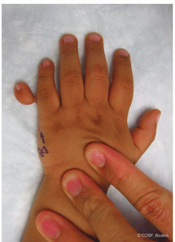 FIGURE 3-1 Photograph of a type B (simple) postaxial polydactyly. |
While simple, the classification of Temtamy and McKusick10 is useful. Type A postaxial polydactyly refers to the well-formed digit that articulates with the fifth (or sixth) metacarpal and has its own set of tendons, neurovascular bundles, and soft tissue structures. Type B polydactyly refers to the rudimentary, underdeveloped digit, which typically is attached to the ulnar border of the hand by only a small skin bridge and neurovascular pedicle.
Stelling11 and Turek12 proposed that postaxial polydactyly be classified into one of three types. Type 1 denotes a rudimentary, hypoplastic digit attached via a small skin bridge. Type 2 postaxial polydactyly refers to a “partial duplication,” in which a normal digit articulates with a bifid or enlarged metacarpal. Type 3 represents the rare complete duplication of metacarpal and phalanges.
Surgical Indications
While some families/patients choose not to treat postaxial polydactyly due to personal, cultural, or religious reasons, in most situations, surgery is recommended to remove the extra digit and provide a more aesthetically normal, functioning hand.
Related posts:

Stay updated, free articles. Join our Telegram channel

Full access? Get Clinical Tree


