Periprosthetic Fractures of the Femur Associated with Hip Arthroplasty
Johannes M. van der Merwe
Donald S. Garbuz
Clive P. Duncan
Bassam A. Masri
Case 1 Scenario
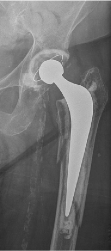
An 84-year-old female presented to the emergency department with hip pain after sustaining a low-energy fall; her hip was previously without symptoms. Radiographs showed a periprosthetic fracture around the stem (Fig. 88.1).
Epidemiology
Periprosthetic fracture of the femur is an uncommon but potentially challenging complication of total hip arthroplasty (THA). The prevalence is rising secondary to the increasing number of hip joint replacements being done worldwide, and the incidence is expected to rise resultant of the advancing age of those who have undergone the procedure and the ever-present issue of osteolysis.
During primary THA, intraoperative periprosthetic fractures have been reported to occur in 0.1% to 3.5% of cases with cemented stems (1,2) and as frequently as 5% of those with cementless stems (3). The latter is due to the greater broaching force that may be required to achieve a stable bone–implant interface and initial implant stability. The risk of intraoperative fractures in the revision setting is even higher with a 3.6% to 6.3% incidence in cemented and 17.6% to 20.9% incidence in uncemented stem revisions (4,5,6).
The true prevalence of postoperative periprosthetic fractures after primary THA is difficult to estimate due to the heterogeneity of the patient population reported in the literature. In the Mayo Joint Registry a prevalence of 1.1% was reported after 23,980 primary THAs (3). This was similar to the findings of Meek and Norwood (7) who found a rate of 0.9% after 5 years in their analysis of 52,136 primary THAs. The Swedish National Hip Arthroplasty Register reported an annual incidence of between 0.045% and 0.13% (8,9). Postoperative fractures have been estimated to occur in approximately 4% of revision THAs (3,7).
Risk Factors
Periprosthetic fractures of the femur are most often associated with low-energy injuries because of a pre-existing decrease in the mechanical strength of the host bone. These risk factors can be divided into systemic and local risk factors.
Systemic risk factors include all causes leading to generalized osteopenia. These include a wide range of disorders ranging from metabolic bone diseases to neuromuscular deficits. Local risk factors focus on poor localized bone stock. These include stress risers, complex deformities of the proximal femur, focal or more widespread osteolysis, bone loss from multiple revision operations, or disuse osteopenia (see Table 88.1). Several surgical techniques and patient
risk factors have been associated with periprosthetic femur fractures. Revision arthroplasty techniques that transfer energy to the tip of the implant stem, for example, impaction allograft and cementless fully porous-coated cylindrical press-fit stems, may predispose the patient to periprosthetic fractures (6,10,11).
risk factors have been associated with periprosthetic femur fractures. Revision arthroplasty techniques that transfer energy to the tip of the implant stem, for example, impaction allograft and cementless fully porous-coated cylindrical press-fit stems, may predispose the patient to periprosthetic fractures (6,10,11).
Table 88.1 Risk Factors Associated with Intraoperative and Postoperative Periprosthetic Femoral Fractures | ||||||||||||||||||||||||||||||||||
|---|---|---|---|---|---|---|---|---|---|---|---|---|---|---|---|---|---|---|---|---|---|---|---|---|---|---|---|---|---|---|---|---|---|---|
|
Based on a biomechanical analysis, increased body mass index (BMI) was associated with an increased fracture risk (12). The Scottish Registry (7) found that the risk of fracture is significantly influenced by the gender and the age of the patient at time of surgery. They found that females (13), patients older than 69 years, and patients undergoing revisions were at an increased risk. These factors may have been confounded by osteoporosis.
Another study (13) found four independent risk factors associated with a significant increased risk of periprosthetic fractures after revision THA. These included female gender, younger age, higher Deyo–Charlson comorbidity index, and an underlying diagnosis of nonunion or fracture. Patients younger than 60 years were at higher risk presumably because they lead a more active lifestyle and are more at risk of traumatic injury. The addition of comorbidities does increase a patient’s risk to sustain a periprosthetic fracture, but it is unclear what the role of optimizing these comorbidities preoperatively might have on the final outcome. They also found that an underlying diagnosis of nonunion or fracture led to a two- to fivefold increased risk compared to patients with only loosening, wear, or osteolysis.
Classification
A functional classification should guide treatment, estimate prognosis, predict likely complications, and permit the meaningful comparison of outcomes between different surgeons or centers (14). The Vancouver classification was developed on the basis of the three most relevant features: fracture location, stem stability, and the quality of the remaining bone stock (15,16). Several studies have demonstrated its validity and reliability (14,17,18).
Intraoperative Fractures
The modified Vancouver classification divides the femur into three anatomic zones (see Table 88.2):
Type A: confined to the trochanteric region without extension into the diaphysis;
Type B: affecting the diaphysis, around or just distal to the tip of the implant;
Type C: involving the diaphysis well distal to the tip of the implant.
Each of these fracture types is then subdivided according to the configuration and stability of the fracture.
Subtype 1 refers to a simple cortical perforation. These fractures typically occur during cement removal from thin cortical bone, or at the time of reaming for canal preparation, and are most common in the diaphyseal region (i.e., type B1).
Subtype 2 is defined as a nondisplaced linear fracture. These fractures typically occur in the proximal metaphysis or diaphysis. It occurs generally at the time of rasp or implant insertion. The increased hoop stress on the bone during impaction is the initiating factor.
Unstable or displaced fracture patterns are termed subtype 3. Fractures occurring in the proximal femur (i.e., type A3) typically occur with forceful impaction of a rasp or the final prosthesis. In the revision setting it can occur when the component is aggressively removed without first clearing the base of the trochanter. Fractures ensuing in the diaphysis
(i.e., type B3) happen commonly with aggressive cement removal, reaming, impaction, or with excessive manipulation of the limb. Fractures appearing in the distal diaphysis (i.e., type C3) rarely occur in isolation and are more commonly associated with the distal propagation of a type B3 fracture. When they do occur in isolation, it can be related to limb rotation in an osteopenic patient.
(i.e., type B3) happen commonly with aggressive cement removal, reaming, impaction, or with excessive manipulation of the limb. Fractures appearing in the distal diaphysis (i.e., type C3) rarely occur in isolation and are more commonly associated with the distal propagation of a type B3 fracture. When they do occur in isolation, it can be related to limb rotation in an osteopenic patient.
Table 88.2 The Modified Vancouver Classification System for Intraoperative Periprosthetic Fractures | ||||||||||||
|---|---|---|---|---|---|---|---|---|---|---|---|---|
|
It is important to understand the difference between an intraoperative B3 (outlined above) and postoperative B3 fracture (outlined below). The former refers to an unstable or displaced fracture during operation and makes no reference to the available bone stock or strength, which are usually adequate. The latter, occurring after operation, means that the available bone stock is poor and complex reconstruction needs consideration.
Postoperative Fractures
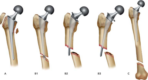
The Vancouver classification integrates the most important factors in the management of postoperative periprosthetic fractures of the femur: fracture location, implant stability, and the quality of femoral bone stock (see Fig. 88.2) (16). The classification system divides the femur into three anatomic regions: (A) apophyses or trochanteric region of the proximal femur; (B) diaphysis, around or just distal to the implant; (C) involving the diaphysis or distal metaphysis well distal to the tip of the prosthesis.
Type A is subclassified as AG, for fractures of the greater trochanter, and AL, for those of the lesser trochanter. Type AG is stable when it is minimally displaced because it is held in place by the digastric effect of the glutei and vasti. Type AL can usually be ignored if occurring in isolation unless it involves the medial cortex and has destabilized the stem with loss of medial support. We describe this type of fracture as a pseudo-type AL (it is in fact a type B2). This fracture pattern destabilizes the stem and needs to be addressed surgically (19,20).
Type B fractures are subclassified depending on the stability of the implant and the available bone stock. This is critical for decision making in treatment.
In B1 fractures the implant is stable. They can be successfully managed with reduction and internal fixation. In type B2 and B3 the implant is loose and stem revision is required. Implant stability while obvious in some cases can be difficult to ascertain; if prior films are available they can be very useful in differentiating well fixed from loose stems. It is important to keep in mind that most periprosthetic fractures are B2 or B3 fractures; B1 fractures are rare and the surgeon should be alert to misclassifying a B2 as a B1 as this is both a common error and will lead to a poor outcome. This was emphasized by Lindahl et al. (21) who demonstrated a 23% failure rate of the treatment of periprosthetic fractures, which was attributed to probable under diagnosis of loose components. It is therefore imperative to scrutinize the preoperative films for signs of a loose stem, and to review the patient history carefully to detect any pain prior to the fracture. If there is any doubt regarding the stability of the stem, the stem should be tested intraoperatively for instability.
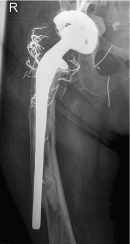
In a B3 periprosthetic fracture, (see Fig. 88.3) the stem is loose and there is severe bone loss such that more specialized techniques of reconstruction may need consideration.
Type C fractures are well distal to the tip of the implant. The modern principles of osteosynthesis are appropriate in these cases, including minimally invasive plate osteosynthesis (MIPO). In general the proximal end of the plate will need to
extend proximal to the tip of the implant to provide adequate fixation. In such cases, modification of the plate attachment will need consideration, such as unicortical screws or cables.
The Vancouver classification system (VCS) has been recently expanded to include two uncommon fracture types:
Type D: Fracture of the diaphysis or distal metaphysis of the femur between both a hip and knee replacement (Fig. 88.4A,B); this pattern has also been referred to as an interprosthetic fracture of the femur.
Type E: Fracture of both the femur and pelvis following hip replacement (Fig. 88.5).
Stay updated, free articles. Join our Telegram channel

Full access? Get Clinical Tree


