Peripheral Compartment Approach to Hip Arthroscopy

CHAPTER 11 Peripheral Compartment Approach to Hip Arthroscopy


Introduction


Arthroscopy without traction of the peripheral compartment (PC) has become an integral part of hip arthroscopy. It has contributed to a better understanding of the functional anatomy and pathogenesis of new concepts such as femoroacetabular impingement and other pathologic conditions of the hip joint. With the tremendous improvement of its technique and the development of better instrumentation, a completely new field of therapeutic options has evolved.


The acetabular labrum is the key structure during portal placement and therapeutic arthroscopy within the hip joint. The labrum seals the joint space between the lunate cartilage and the femoral head by maintaining a vacuum force of about 120 N to 200 N, which keeps the femoral head within the socket. To overcome the vacuum force and the passive resistance of the soft tissues, traction is needed to separate the head from the socket, to elevate the labrum from the head, and to allow the arthroscope and other instruments access to the narrow “artificial space” between the weight-bearing cartilage of the femoral head and the acetabulum. However, if traction is applied, the joint capsule with the iliofemoral, ischiofemoral, and pubofemoral ligaments is tensioned, and the joint space peripheral to the acetabular labrum decreases. Thus, to maintain the space of the PC for better visibility and maneuverability during arthroscopy, traction should be avoided.


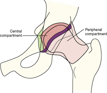
The hip is divided arthroscopically into two compartments that are separated by the labrum (Figure 11-1). The first is the central compartment (CC), which comprises the lunate cartilage; the acetabular fossa; the ligamentum teres; and the loaded articular surface of the femoral head. This part of the joint can be visualized almost exclusively with traction. The second is the PC, which consists of the unloaded cartilage of the femoral head; the femoral neck, with the medial, anterior, and posterolateral synovial folds (i.e., Weitbrecht ligaments); and the articular capsule with its intrinsic ligaments, including the zona orbicularis. This area can be better seen without traction.


In addition to its importance in therapeutic hip arthroscopy, the PC can be used as an intermediate space to control the placement of portals to the CC. In our practice, we have been using this technique successfully for more than 4 years; it has a smaller risk of iatrogenic lesions of the acetabular labrum and the femoral head cartilage.


Imaging and diagnostic studies


Radiologists and surgeons need to consider that the hip joint is not limited to the direct articulation between the cartilage of the femoral head and the lunate cartilage of the acetabulum. The PC joint extends to the intertrochanteric line anteriorly and to the femoral neck posteriorly. Thus, the assessment of roentgenograms, computed tomography scans, and magnetic resonance images has to include this part of the joint.


Consequently, radiographs need to be evaluated for the contour of the head–neck junction, the overcoverage of the anterolateral acetabular rim, and perilabral ossifications in cases that involve femoroacetabular impingement. Here, one has to differ between the primary signs of impingement (i.e., loss of offset) and osteophytes, which are signs of osteoarthritis and secondary changes. In general, the removal of osteophytes does not lead to the same good results as the reshaping of the head–neck junction in femoroacetabular cam impingement. In addition, osteophytes are indicators of cartilage degeneration within the CC, which is important for the patient’s prognosis. The soft tissues around the femoral neck have to be analyzed for subtle or more prominent ossifications that indicate osteochondromas within the PC. With magnetic resonance imaging, the space underneath the articular capsule needs to be scanned for an effusion; inflammation; thickening; villous or nodular hypertrophy of the synovium; loose bodies; and chondromas. The latter often accumulate in the pouch underneath the transverse ligament and in the gutter medially to the femoral neck. Depending on their cartilaginous or bony structure, size, and location, one has to decide whether to perform the removal through the arthroscope or via open surgery. The labrum needs to be analyzed for the degree of degeneration, tears at the labrum–cartilage junction, intralabral or perilabral cysts, and ossifications. These are important indicators for determining whether the labrum can be saved, trimmed or resected, or temporarily detached for the trimming of the bony rim. Some of this intra-articular work can be done under traction from the CC, and some can be done without traction via the PC. Radial magnetic resonance cuts can be helpful for the better imaging of the contour of the head–neck junction in different positions. For cases in which the loss of offset extends posterolaterally, only very experienced arthroscopic surgeons are capable of reshaping the head–neck junction sufficiently.


Diagnostic injections of the hip joint with local anesthetics are very helpful for the differentiation of intra-articular causes of pain from those that are extra-articular. Under fluoroscopy or ultrasound, the needle is introduced via the proximal anterolateral portal to the anterolateral head–neck junction and into the PC. Here, the space between the capsule and the bone allows for safe and effective fluid aspiration or injection. The straight anterior puncture or injection without imaging has a high risk of periarticular misplacement.


Surgical technique


Positioning and Switching Between the Compartments


Hip arthroscopy with and without traction can be performed with the patient in the lateral or supine position. From our experience, the decision of whether to use the lateral or supine position is more a matter of individual training and habit of use. However, because of the almost exclusive use of the proximal and distal anterolateral and anterior portals during hip arthroscopy without traction, we prefer the supine position.


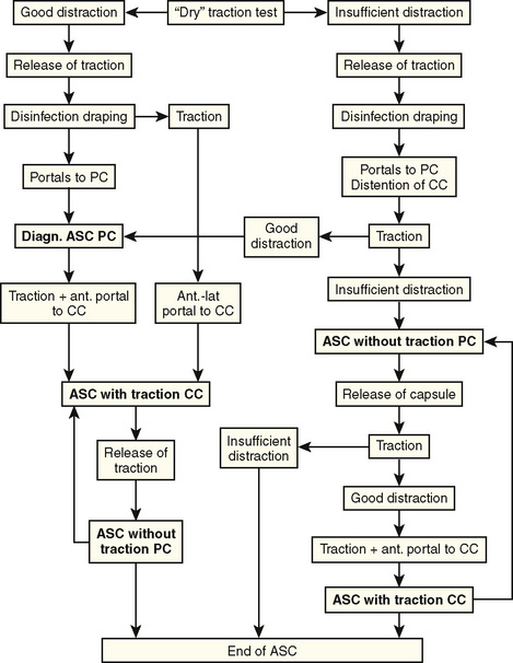
To allow for a complete diagnostic arthroscopic examination of the hip, both techniques with and without traction are combined for arthroscopy of the CC and the PC; this requires specific attention to positioning, table equipment, and draping. The order of arthroscopy with and without traction depends on different parameters (Figure 11-2). In “standard” cases with good distraction and visibility, we prefer to access the PC first to control portal placement to the CC. However, there are cases in which the distraction of the hip is insufficient and visibility in the PC decreased. Here, a release of different parts of the joint capsule can be performed to increase the PC space and improve distraction. We usually start with a release of the zona orbicularis that extends into the iliofemoral ligament in cases of severe capsular thickening or fibrosis, and this is followed by therapeutic procedures such as the reshaping of the head–neck junction. At this point, traction is again applied. If distraction is improved, portals to the CC can be placed under arthroscopic control. However, if distraction is still not sufficient, arthroscopy of the PC only should be considered to avoid iatrogenic damage of the acetabular labrum and the femoral head cartilage during portal placement to the CC.


For arthroscopy of the PC, a good range of movement is important to relax parts of the capsule and to increase the intra-articular volume of the area that needs to be inspected and addressed. In addition, only without traction can the impingement maneuver and other functional tests be reproduced under various degrees of flexion, rotation, and abduction. Another advantage of the nontraction technique is the possibility of “hiding” the hyaline cartilage of the femoral head under the socket by flexion to avoid damage when working in the PC.


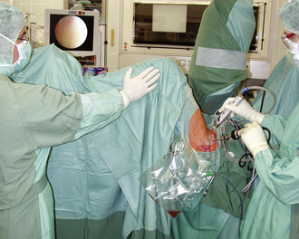
During the past several years, we have removed the foot from the traction module to allow for the maximum range of movement. However, this technique is time consuming and strenuous for the assistant who is holding the leg. In addition, switching back into the CC under traction is difficult, because the foot needs to be readapted to the traction module. Especially for femoroacetabular impingement cases, there is a need to alternate between both compartments with and without traction. Thus, we changed our technique. For arthroscopy of the PC, the foot is kept in the traction module. The traction is released, and the traction module with the foot is slid in with the extension bar. With this technique, the hip and knee can be flexed up to 90 degrees, abducted to about 30 degrees, and rotated 20 degrees internally and externally (Figure 11-3). When reentering the CC, the extension boom is drawn out, and the hip and knee are extended. Before traction is applied, the room nurse has to check for possible soft-tissue entrapment at the perineum.


Portals to the Peripheral Compartment


Basically, portals are established where they are needed. With our current knowledge, most pathology in the PC can be addressed via anterior and lateral portals. It is still difficult to access the posteromedial parts of the PC for both viewing and working. The posteromedial peripheral space is significantly smaller and additionally hidden from the posterior part of the greater trochanter. Thus, this part of the PC can sometimes be better seen and accessed via the CC under traction. The distal part of the posteromedial space of the PC cannot be seen from the CC but rather from the perilabral sulcus and the space directly peripheral to the posterior part of the transverse ligament. Here, loose bodies and chondromas can be reached via the posterolateral portal.


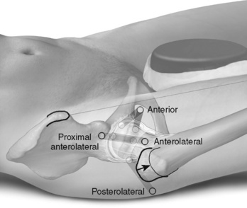
A comprehensive overview of the PC can be obtained from the proximal anterolateral portal only (Figure 11-4). At this level, the soft-tissue mantle is relatively thin, and the position of the portal is near the lateral cortex of the femoral head. Therefore, the maneuverability of the arthroscope is sufficient for moving it into the medial recess, gliding over the anterior surface of the femoral head to the lateral recess, and passing the lateral cortex of the femoral neck for the inspection of the posterolateral recess. With the need for better access to the lateral and posterolateral head and neck area for therapy of the cam type of femoroacetabular impingement and the removal of chondromas from the medial area underneath the transverse ligament in patients with synovial chondromatosis, a two-portal technique for the PC is usually preferred.

< div class='tao-gold-member'>

Stay updated, free articles. Join our Telegram channel

Jul 24, 2016 | Posted by in MUSCULOSKELETAL MEDICINE | Comments Off on Peripheral Compartment Approach to Hip Arthroscopy

Full access? Get Clinical Tree

Get Clinical Tree app for offline access