CHAPTER 37 Pediatric Pelvic Osteotomies and Shelf Procedures
Introduction
The goal of the treatment of a child with developmental dysplasia of the hip is to attain a radiographically confirmed normal hip at maturity to hopefully prevent degenerative joint disease. Residual acetabular dysplasia, with or without hip subluxation, will lead to degenerative joint disease. Studies of the causes of degenerative joint disease of the hip estimate that 20% to 50% of cases may be attributed to acetabular dysplasia or hip subluxation, especially among females. The correction of residual acetabular dysplasia theoretically provides for a better weight-bearing surface for the femoral head, restores normal biomechanics of the hip, reduces contact pressures, and increases the longevity of the hip. Biomechanical studies have shown that the onset of joint degeneration correlates with both the magnitude and duration of exposure to contact stresses above 2 MPa.
Basic science
The normal growth and development of the hip joint require a genetically determined balance of growth of the acetabular and triradiate cartilages in conjunction with a well-located femoral head. By the eleventh week of intrauterine life, the hip joint is fully formed. Several factors come into play to allow for the normal development of the acetabulum. The main stimulus for the concave shape of the acetabulum is the presence and maintenance of a reduced spheric femoral head. There must also be normal interstitial and appositional growth within the acetabular cartilage as well as periosteal new bone formation in the adjacent bones of the pelvis. At puberty, the development of three secondary centers of ossification serves to further enhance the depth of the acetabulum. The os acetabulum is the epiphysis of the pubis, and it forms the anterior wall of the acetabulum. The acetabular epiphysis is the epiphysis of the ilium, and it forms the superior edge of the acetabulum. The ischial epiphysis also contributes to normal growth.
In a child with developmental hip dysplasia, some aspects of normal growth and development are altered. The femoral head is the key stimulus for acetabular development, so it must be reduced as soon as possible. If the reduction is maintained, it will provide the stimulus for acetabular development, and there is the potential for the recovery and resumption of normal growth and development. The capacity for the acetabular cartilage to resume normal growth depends on its intrinsic growth potential and on whether it was damaged by the subluxated or dislocated femoral head, by various attempts at reduction, or by surgery. After the age of 4 years, the potential for the restoration of normal anatomy is markedly decreased.
Indications
History and physical examination
Children with residual dysplasia are most often asymptomatic. Early complaints may be of vague discomfort with activity; complaints of more severe pain may indicate the presence of degenerative changes in the hip. Catching, locking, or giving way may be indicative of labral pathology. Sharp groin pain, especially with hip flexion, may suggest acetabular impingement.
The physical examination should include an assessment of the hip’s range of motion, joint contractures, motor strength (specifically of the abductor), limb alignment, limb-length discrepancy, and gait pattern. Physical findings are usually normal, even among patients with severe dysplasia. With significant symptoms, however, a limp or an abductor lurch may be evident. Pelvic obliquity is assessed by palpating the posterior iliac crests while the patient is standing. Patients with acetabular impingement may have decreased motion or pain with the provocative maneuvers of hip flexion and internal rotation.
Imaging studies
The evaluation of dysplasia can be accomplished in most cases with the use of plain radiographs. Essential views include a standing anteroposterior pelvic view and a frog-leg lateral view. The anteroposterior view allows for the evaluation of the Shenton line; a broken Shenton line indicates subluxation of the hip. The acetabular sourcil (which is French for “eyebrow”) is usually a smooth curve of uniform thickness. In dysplastic hips, the sourcil is thicker laterally, which indicates a focal loading of the joint; it may also fail to completely “turn down” at its peripheral extent. On the anteroposterior view, measures of the acetabular index, the center-edge angle of Wiberg, and the acetabular angle of Sharp can also be made. The abduction and internal rotation view can be used to assess the true femoral neck–shaft angle and to simulate the amount of coverage possible with proximal femoral derotation osteotomy. The false-profile view is a true lateral view of the acetabulum, and it allows for the assessment of the anterior coverage of the femoral head. Three-dimensional computed tomography scans may provide increased information regarding the shape and orientation of the acetabulum and of the fit of the femoral head within it. Magnetic resonance imaging may be useful to evaluate any underlying labral pathology.
Surgical techniques
Treatment options are divided into three groups: rotational osteotomies, volume-reducing osteotomies, and salvage procedures.
Rotational Osteotomies
Rotational osteotomies attain femoral head coverage by cutting one to three of the pelvic bones, with the acetabulum being rotated on the intact structures. These osteotomies cover the femoral head with acetabular cartilage, and they intuitively are the first choice for femoral head coverage procedures. They all require a concentrically reduced femoral head.
The innominate osteotomy of Salter is the most widely used of the rotational osteotomies for the pediatric population. It divides the ilium just above the acetabulum, which allows the acetabulum to be rotated through the symphysis pubis. Neither the contour nor the volume of the acetabulum is changed. This procedure can obtain 20 degrees of improvement in the center-edge angle and 10 degrees of improvement in the acetabular index.
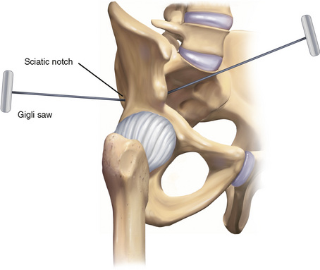
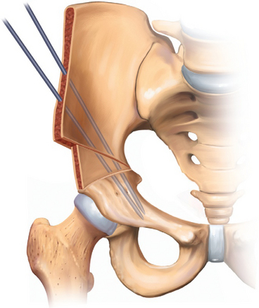
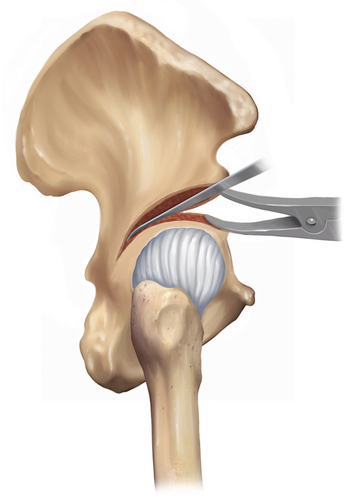
The patient is placed in the supine position with a sandbag under the ipsilateral thorax, and the affected limb is draped free. Adductor contracture is released with a subcutaneous or open tenotomy. Incision and exposure are provided via a Smith-Peterson approach to the hip. A so-called bikini incision starts 2 cm distal to the center of the iliac crest, extends 1 cm distal to the anterosuperior iliac spine, and ends below the middle of the inguinal ligament. The interval between the tensor fascia latae and the sartorius is developed to expose the rectus femoris and the anteroinferior iliac spine. The iliac apophysis is incised down to bone along the iliac crest from the posterior end of the skin incision to the anteroinferior iliac spine. The lateral portion of the apophysis and the periosteum of the outer table are carefully stripped in a continuous sheet to the lateral edge of the acetabulum and posteriorly to the greater sciatic notch; this space is then packed. Adhesions of the joint capsule to the lateral aspect of the ilium can be freed with a periosteal elevator. The medial half of the apophysis and the periosteum of the inner wall are carefully stripped in a continuous sheet to expose the sciatic notch. Care is taken to remain subperiosteal to avoid injury to the sciatic nerve and the superior gluteal artery. The tendinous portion of the iliopsoas is exposed on its deep surface at the level of the pelvic brim and rolled over to visualize the musculotendinous junction. A scissors is passed between the tendon and the musculotendinous junction, and the tendon is cut sharply with a scalpel. The tip of a curved forceps is passed subperiosteally from the medial side and through the sciatic notch to grasp the end of the Gigli saw. The index finger of the opposite hand is used to guide the forceps. The skin and the soft tissues are retracted widely. The osteotomy extends in a straight line from the sciatic notch to the anteroinferior iliac spine, with care taken to remain at right angles to the vertical axis of the ilium. The hands are kept far apart, and continuous tension is placed on each end of the saw to keep it from binding (Figure 37-1). A triangular-shaped bone graft is then taken from the iliac crest with large bone cutters. (A saw may be used in older children.) The base of the graft extends from the anterosuperior iliac spine to the anteroinferior iliac spine. A towel clip is placed on each fragment. The proximal fragment should only be steadied. The towel clip on the distal fragment should be placed well posterior to avoid fracture. The distal fragment is then rotated downward and forward in line with the ilium, which opens the osteotomy anterolaterally. Avoid the posterior or medial displacement of the distal fragment. If the hip capsule has not been opened, the leg can be used to attain the desired correction by placing it in a figure-four position. Downward pressure on the knee as the heel is moved toward the child’s chin produces the desired rotation. The wedge-shaped bone graft is then inserted into the osteotomy site, and the traction is released from the distal fragment. The posterior aspect of the osteotomy should remain closed. Two heavy, threaded K-wires are inserted across the osteotomy site, through the graft, and into the distal segment that lies medial and posterior to the acetabulum (Figure 37-2). Care is taken to avoid penetrating the hip joint. The hip is carefully moved so that crepitus can be heard and felt for; this may indicate that a pin has penetrated the joint. The two halves of the iliac apophysis are sutured together. The K-wires are cut so that their ends lie in subcutaneous fat. A drain is usually not necessary if only an innominate osteotomy has been performed. The wound is then closed. A 1½ hip spica cast is applied with the hip in slight abduction, flexion, and internal rotation and with the knee in flexion. In older, reliable children, three-point partial weight bearing with crutches may be permitted with no immobilization.

Figure 37–1 The inner and outer tables of the ilium are exposed. The periosteum is elevated carefully from the sciatic notch with a curved periosteal elevator, such as a Crego elevator. A right-angled forceps is passed medial to lateral while the finger of the other hand is used to push the periosteum down and away from the sciatic notch on the outer table. A Gigli saw is grasped in the forceps and pulled through the sciatic notch. Retractors are placed in the sciatic notch on each side of the ilium to provide wide retraction and to protect the soft tissues. The osteotomy is performed with the Gigli saw, which emerges at or just above the anterior inferior iliac spine. While using the Gigli saw, the hands should be spread as far apart as possible. Constant tension should be kept on each end of the Gigli saw, because it has a tendency to bind. The bone graft that is used to hold the osteotomy site open is taken from the anterior iliac crest. This can be done with bone-biting forceps in young children, but it is facilitated by the use of a power saw in older children. At this point, it is imperative to perform an intramuscular tenotomy of the iliopsoas as decribed for the anterior approach to the congenitally dislocated hip.

Figure 37–2 Although the graft should be secure, it is not secure enough at this point to be left alone without fixation. Smooth or thin wires should not be used. Two heavy threaded Kirchner wires should be used and passed from the proximal fragment into the distal fragment. In the distal fragment, these should lie medial and posterior to the acetabulum; this determines their starting point in the proximal fragment. There is a danger of passing one of the wires into the hip joint when the capsule has not been opened, as may occur during the treatment of acetabular dysplasia. This danger and the fact that properly placed pins appear on the postoperative radiograph to be penetrating the hip joint make it imperative that the surgeon has a good grasp of the pelvic anatomy and that he or she carefully moves the hip to feel and listen for crepitus. After this, the wound is closed. A drain is usually not necessary when only an innominate osteotomy has been performed.
Osteotomies in which all three pelvic bones (e.g., Steel, Tönnis, Carlioz) are cut offer greater rotational advantages. The Ganz periacetabular osteotomy may be performed for patients with a closed triradiate cartilage. This procedure is discussed in detail in Chapter 26, and it will not be discussed here. Variations of the triple osteotomy exist for patients with open triradiate cartilage.
Volume-Reducing Osteotomies
Volume-reducing osteotomies involve the use of incomplete cuts and hinge on different aspects of the triradiate cartilage; thus, these procedures are limited to patients with open triradiate cartilage.
The Pemberton osteotomy addresses a size “mismatch” between the acetabulum and the femoral head. By hinging on the posterior limb of the triradiate cartilage, the shape of the acetabulum can be changed to provide improved coverage for the femoral head.
The patient is positioned supine, with a bump placed under the involved hip. Exposure is via a Smith-Peterson approach to the hip. Just as with the Salter osteotomy, the iliac apophysis is split, and both the inner and outer tables of the ilium are exposed subperiosteally. Exposure is carried out to the sciatic notch, and the rectus insertion is left alone. Although it was not recommended by Pemberton in his original article, the division of the psoas tendon (as in the Salter osteotomy) may facilitate correction. Two flat-blade retractors are inserted into the sciatic notch on either side of the ilium. The osteotomy is performed with a narrow curved osteotome through the outer table, starting 1 cm above the anteroinferior iliac spine and extending posteriorly, keeping 1 cm to 1.5 cm from the attachment of the hip capsule. Because the osteotomy is carried posteriorly and then inferiorly through the outer table, it will disappear into the soft-tissue attachments behind the capsule. Visualization can be facilitated by the rotation of the retractor in the sciatic notch. Care must be taken to avoid cutting into the sciatic notch. Direct the osteotomy to the ilioischial limb of the triradiate cartilage. With the use of the same osteotome, a corresponding cut is made on the inner table. As with the outer table, avoid cutting into the sciatic notch. The plane of the osteotomy may be adjusted on the basis of the type of coverage that is necessary. A more transverse cut will provide more anterior coverage, whereas a laterally inclined osteotomy will provide more lateral coverage. After both cortices of the ilium have been cut, a wide curved osteotomy is used to connect the two cuts. As the osteotome proceeds posteriorly, it will become apparent that it cannot make the sharp turn inferiorly into the posterior column. A special Pemberton right-angled curved osteotome is used to complete this cut into the triradiate cartilage (Figure 37-3). A small lamina spreader can hold the osteotomy apart and facilitate this cut. The acetabulum can now be directed into the desired position. A groove is cut into each surface of the osteotomy with a narrow gouge or curette. A triangular wedge of bone is removed from the anterior iliac crest and placed in the osteotomy site (Figure 37-4). Because the graft will be recessed in the osteotomy site, the graft should be larger than the gap created by the osteotomy. This osteotomy is quite secure, and it does not require additional fixation. The iliac apophysis is reapproximated, and the wound is closed. The patient is placed in a 1½ hip spica cast.

Figure 37–3 After the inner and outer cortices of the ilium are divided as far as can be seen, a wider curved osteotome is used to connect these two cuts. As the osteotome proceeds posteriorly, it becomes apparent that it is not able to make the sharp turn inferiorly to avoid cutting into the sciatic notch. At this point, an osteotome with a right-angled curve (available via special order from Zimmer Co., Warsaw, IN) is inserted into the osteotomy. This can be made easier by prying down on the acetabular roof with an osteotome and then inserting a small lamina spreader to hold the osteotomy apart. The special osteotome is used to complete the cut into the triradiate cartilage. It is not possible to see the tip of the osteotome as it completes the osteotomy. It is not necessary and usually not possible to see the triradiate cartilage unless the acetabulum is levered down excessively. When the osteotomy is complete, the acetabular roof can be levered down into the desired position and held there with a lamina spreader.

Stay updated, free articles. Join our Telegram channel

Full access? Get Clinical Tree

