Chapter 96 Osteotomy About the Knee
International Roundtable Discussion
Osteotomy about the knee has traditionally been used to treat the middle-aged patient with gonarthrosis, but the indications have broadened to include the younger patient or athlete with ligament deficiencies, chondral defects, absent menisci, or posttraumatic degenerative arthritis. We have assembled a panel of experts that includes Michael Stuart (moderator), David Backstein, Martin Logan, and Thomas Muellner, who will share their personal ideas and preferences based on extensive experience with this procedure.
Michael Stuart
For many orthopedic surgeons, unicompartmental or total knee arthroplasty has supplanted realignment osteotomy as their primary treatment for gonarthrosis.
David Backstein
The frequency of osteotomy for unicompartmental arthritis of the knee has declined somewhat in my practice over the past 5 to 10 years; however, I strongly believe that there are still several excellent indications for osteotomy around the knee. In addition to unicompartmental arthritis, other indications include osteonecrosis, adult osteochondritis dissecans, and osteotomy in combination with osteochondral grafting to off-load the involved compartment.
Candidates must have adequate bone stock to allow effective fixation and early range of motion, a preoperative range of motion arc of at least 90 degrees, and less than 15 degrees flexion-contracture. Because of osteoporosis and its impact on fixation, it is my practice to avoid performing an osteotomy on males older than 65 years and females older than 60 years.
Martin Logan
Undoubtedly, osteotomy has a role in patients with isolated unicompartmental osteoarthritis. Our improved understanding of knee kinematics and joint implant design makes unicompartmental arthroplasty a more attractive option than ever before. That said, the joint registry data on unicompartmental arthroplasty around the world show a higher failure rate in patients younger than 55 years of age.
Factors that affect decision making include patient motivation, understanding, compliance, and occupation. For example, a manual laborer in his mid 40s is a better candidate for osteotomy rather than unicompartmental arthroplasty. A sedentary female with isolated medial osteoarthritis and normal preoperative alignment is unlikely to be satisfied functionally and cosmetically with an osteotomy; therefore, I would prefer a unicompartmental arthroplasty.
Michael Stuart
We all agree that osteotomy still has a role in selected patients with unicompartmental arthritis and malalignment.
Thomas Muellner
There is absolutely a role for realignment osteotomies in treating isolated medial or lateral compartment arthritis in patients with varus or valgus malalignment. In my practice, the ideal candidate for a realignment procedure is the middle-aged patient who has pain during heavy work or recreational sports activities. If instability is an accompanying symptom, combined realignment and ligament reconstruction should be considered.
David Backstein
Yes, the ideal candidate for a proximal tibial valgus osteotomy is a young, physically active patient with medial compartment osteoarthritis of the knee and varus tibiofemoral alignment, for example, the young patient with a highly physical job such as a firefighter or construction worker, who possesses the capacity for a fairly prolonged and at times arduous rehabilitation process. Although this is not commonly encountered, young and active individuals with isolated lateral compartment arthritis and valgus alignment are similarly excellent candidates for osteotomy and are treated with distal femoral varus osteotomy.
Martin Logan
In my mind, the ideal candidate is a male in his early 50s with normal BMI—a highly motivated nonsmoker who has isolated medial osteoarthritis with normal patellofemoral articulation, intact lateral meniscus, normal cruciate ligaments, and a preserved lateral tibiofemoral joint. The knee would be in 5 to 10 degrees of varus compared with the normal contralateral limb and would have a full range of motion without a fixed flexion deformity.
Michael Stuart
The consensus seems to be that an osteotomy is a good option for the young, active patient with a stable, arthritic knee and good bone stock.
David Backstein
Contraindications for a proximal tibial valgus osteotomy include moderate to severe lateral compartment arthritis or significant and symptomatic patellofemoral arthritis. In particular, I avoid an osteotomy if the patient experiences anterior knee pain when climbing or descending stairs, after prolonged sitting, or when getting up from a chair. Osteotomy is also contraindicated in patients with an arc of motion less than 90 degrees, a flexion deformity greater than 15 degrees, and maximum flexion less than 90 to 100 degrees. Inflammatory arthritis, which by its very nature affects the entire joint in a congruous manner, is considered unacceptable for this operation. Two additional situations that are relative contraindications to osteotomy include (1) a high adductor moment (varus thrust), because it is associated with poorer results and recurrence of varus deformity after tibial osteotomy; and (2) obesity, which has been shown to be a risk factor for early failure of high tibial osteotomy.
Martin Logan
Absolute contraindications in my practice include inflammatory arthropathy, previous meniscectomy or arthritis in the compartment intended for weight bearing, gross obesity with BMI over 50, and a patient who is a smoker or a noncompliant patient.
Thomas Muellner
I would add the uncooperative patient, loss of bone with “teeter effect,” and an associated hip flexion contracture.
Michael Stuart
The orthopedic literature has taught us that the success of an osteotomy is dependent on adequate correction of limb malalignment (see Coventry M, Ilstrup D, Wallrichs S: Proximal tibial osteotomy: a critical long-term study of eighty-seven cases. J Bone Joint Surg Am 75:196–201, 1993). Therefore, it is essential to accurately determine the desired amount of correction before surgery and then employ meticulous surgical technique.
Martin Logan
I always get long-leg weight-bearing films and use the Miniaci technique, usually aiming to overcorrect by around 2 degrees and taking into consideration the natural alignment of the contralateral limb (see Miniaci A, Ballmer FT, Ballmer PM, Jakob RP: Proximal tibial osteotomy: a new fixation device. Clin Orthop Relat Res 246:250–259, 1989).
David Backstein
I also get full-length anterior-posterior weight-bearing radiographs of the lower limbs, including the hips, ankles, and knees, to establish the mechanical axis, the anatomic axis, and the point of intersection of the weight-bearing line at the joint line. I aim for a correction that results in passage of the weight-bearing line through the 62% coordinate of the tibia articular surface (medial border of tibia articular surface is 0%, and lateral border is 100%), resulting in preferential loading of the lateral tibiofemoral compartment. The angular correction is calculated by drawing a line from the center of the femoral head to the 62% coordinate of the tibia at the knee. A second line is then drawn from the center of the ankle to the 62% coordinate. The angle between the first and second lines represents the angle of correction required.
I don’t use gait analysis: however, I do examine the patient’s gait, and I consider a severe varus thrust as a relative contraindication to HTO.
Thomas Muellner
In patients who are potential candidates for a realignment procedure, it is essential to assess the alignment on long-standing radiographs (femorotibial angle, LDFA, MPTA, Mikulicz’s line), lateral radiographs of the knee in 90 degrees of flexion, patella tangential, and MRI of the knee. I also use intraoperative control with the image intensifier, which allows accuracy of about ±2 degrees.
Unfortunately, I have only restricted access for gait analysis in my patients to study the adduction moments.
Michael Stuart
I don’t have any experience with osteotomy computer navigation, but it may be helpful for intraoperative verification of multiplanar corrections in complex cases. I routinely use the weight-bearing line method because it is a simple and reproducible technique for determining the desired coronal plane correction angle (Fig. 96-1) (see Dugdale TW, Noyes FR, Styer D: Preoperative planning for high tibial osteotomy: the effect of lateral tibiofemoral separation and tibiofemoral length. Clin Orthop Relat Res 274:248–264, 1992). The surgeon chooses the desired coordinate, which equals the angle of correction according to the specific clinical situation (typically 62%; range, 50% to 75%). Remember that standing radiographs can overestimate the magnitude of correction as the result of osseous defects and/or attenuated ligaments. Compare the amount of lateral joint space opening (in millimeters) with the contralateral knee and subtract the difference from the calculated angle (1 degree per millimeter) to avoid overcorrection.

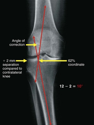
Figure 96-1 The weight-bearing line method is a simple technique for determining the coronal plane correction angle.
(From Stuart MJ: Opening wedge-proximal tibial osteotomy. In Lotke PA, Lonner JH [eds]: Knee arthroplasty, ed 3, Baltimore, 2009, Wolters Kluwer Health/Lippincott William & Wilkins [Master Techniques in Orthopaedic Surgery], pp 361–371.)
The lateral closing wedge osteotomy of the proximal tibia was the procedure of choice in the past to correct a varus deformity. In recent years with the advent of new implant designs, the medial opening wedge technique has become popular.
David Backstein
My preference for varus deformity correction of less than 15 degrees is a medial, opening wedge osteotomy. For situations in which the deformity is 15 to 20 degrees, I perform a closing wedge lateral osteotomy because of the lesser risk of nonunion by opposing and compressing host bone. A fixed varus deformity greater than 20 degrees may necessitate an osteotomy of both the proximal tibia and the distal femur to achieve correction.
Pearls
Thomas Muellner
In patients with an extra-articular tibial varus deformity, I prefer an opening wedge osteotomy. In patients with a combined deformity of the distal femur and the proximal tibia, combined osteotomies have to be considered to avoid an oblique joint line.
Pearls

Stay updated, free articles. Join our Telegram channel

Full access? Get Clinical Tree

