Osteochondritis Dissecans of the Elbow


Osteochondritis Dissecans of the Elbow


Scott A. Tucker

Felix H. Savoie III

Michael J. O’Brien


INTRODUCTION

First described by Konig (1) in 1889, osteochondritis dissecans (OCD) is a localized condition of the articular cartilage and subchondral bone resulting in segmentation and separation from the surrounding bone. The term “osteochondritis dissecans” is defined as an inflammatory condition that causes localized necrosis and fragmentation; however, no inflammatory histology has been elucidated (2). While the original description by Konig (1) resulted from loose bodies within the hip and knee, OCD has also been found within the elbow, most commonly affecting the central and anterolateral capitellum, but also reported in the olecranon, radial head, and trochlea. OCD of the capitellum is not to be confused with Panner disease, which is osteochondrosis of the capitellar ossification center (1,2,3,4 and 5). Panner disease is a benign, self-limited disorder that occurs in younger patients with no apparent sequela. The exact etiology still remains unclear, but recent research suggests that repetitive microtrauma may play a role. There is also increasing evidence that ischemia from a tenuous capitellar epiphyseal blood supply and minimal metaphyseal vascular contribution could predispose the capitellum to avascular necrosis (6). Increased pressure from an inflamed posterolateral plica has also been postulated as a source of pain, restricted motion, and progression of the disorder by increasing the pressure on the dysvascular capitellum.

Elbow OCD generally afflicts athletes in the second decade, particularly those involved in repetitive elbow overuse, such as gymnasts, pitchers, and swimmers. It more commonly affects male athletes, primarily the dominant arm in throwers or overhead athletes, but has been found to be bilateral in 5% to 20% of patients (7,8).

Patients generally present with an insidious onset of vague elbow pain that may localize to painful swelling of the lateral elbow, particularly with increased activity and noticeable loss of motion. Occasionally, patients may complain of mechanical symptoms, such as popping, clicking, or instability with load-bearing activities. The classic physical findings include swelling of the normal posterolateral elbow plica, popping and locking as the inflamed plica is pinched between the radial head and capitellum during range of motion, and loss of terminal extension. Unlike many throwing athletes with medial-sided elbow pathology, and unique to the diagnosis of capitellar OCD, the patient will generally have lateral-sided elbow pain with moving valgus stress testing, though both medial and lateral-sided pathologies may be present.

Imaging studies begin with standard elbow radiographs, which often illustrate the classic findings of central lucency of the capitellum (Fig. 16-1). In later stages, there may be increased bony sclerosis in the center of this lucency and possibly loose bodies. Magnetic resonance imaging (MRI) has become the standard modality for advanced evaluation of these lesions, and specific attention should be paid toward the bony involvement, loose bodies, and the integrity of the overlying cartilage cap. Various MRI sequences can be indicative of disease staging. Early lesions will demonstrate changes on T1, but not on T2 sequences, and the cartilage cap will appear intact. Advanced lesions will show changes on both MRI sequences, as well as possibly loose in situ fragments or frank loose bodies. Advancement of the lesion is further demonstrated by synovial fluid layering between the lesion and remaining capitellum, and most severe cases will display lesions that advance to the lateral edge or
“shoulder”; these lesions often require more reconstructive procedures, so three-dimensional imaging is helpful to assess this integral lateral cortex (Fig. 16-2).


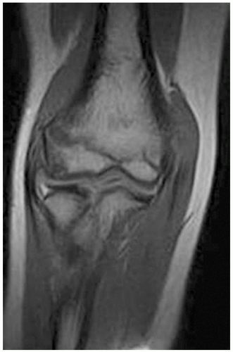
FIGURE 16-1 This MRI demonstrates an early capitellar OCD lesion, as noted by the destruction of the cartilage cap, but no evidence of ring enhancement of the lesion.

Bradley and Petrie classified lesions based on radiography and MRI. For type IA “very early lesions,” radiographs are normal, and the capitellar viability is high. Often, these lesions do not require MRI with contrast; these lesions generally resolve with nonoperative management and close follow-up. Type IB “early lesions” have the more classic radiographic findings of capitellar sclerosis or flattening and are more at risk of cartilage breakdown and fragment instability; thus, intravenous and intra-articular contrast provide the most benefit to assess cartilage integrity and
fragment viability (Fig. 16-1). Dye leaking between the fragment and surrounding bone indicates a break in the cartilage, and therefore, operative stabilization or excision is required. Type II lesions demonstrate more advanced disease on radiographs, such as sclerosis surrounding the lesion, and the patient may complain of mechanical symptoms from a loosely attached lesion (Fig. 16-2). Type II lesions are differentiated from type III lesions by the chronicity and presence of intra-articular loose bodies, though both types normally warrant operative treatment to stabilize and/or debride the lesion. Lastly, Bradley and Petrie (5) described the rare type IV lesions as those with a “bipolar” lesion noted on the radial head, which can be simultaneously treated with debridement, curettage, and subchondral drilling at the time of capitellum surgery.


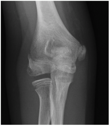
FIGURE 16-2 AP radiograph demonstrating an advanced capitellar OCD lesion, as noted by the decreased capitellar density and perifragmentary sclerosis.


INDICATIONS/CONTRAINDICATIONS

Indications for surgical management are generally limited to patients who have failed a lengthy period of nonoperative management, and, as in most cases, surgical intervention should be reserved for patients with pain and/or functional impairment. Specific attention should be made to the status of the cartilage cap on radiographs or MRI, as absence of an intact cartilage cap is more likely to require surgery. Radiographic progression of the disease process, evidence of symptomatic loose bodies, and cartilage cap disruption that persists despite rest, bracing, and activity cessation are all indications to proceed to surgery. Baumgarten et al. specifically proposed a classification system for OCD lesions (Table 16-1). This study recommends drilling for grade 1 lesions, cartilage debridement to a stable rim and abrasion chondroplasty of subchondral bone for grade 2 lesions, fragment removal and abrasion chondroplasty for grades 3 and 4, and removal of loose bodies with similar treatment for the bony bed in grade 5 lesions (9). Recent reports also have delineated worse results with those lesions involving the lateral aspect of the capitellum. Thus, lesions may also be classified as constrained and having an intact lateral shoulder or unconstrained with an involvement and destruction of the lateral aspect with resultant worse prognosis (10,11).

Contraindications to surgery include those patients with intact articular cartilage. Patients who have not yet exhausted or have responded favorably to conservative management, such as rest, bracing, and cessation of aggravating activities, should not yet undergo surgical intervention.


TABLE 16-1 The Arthroscopic Classification System of the Elbow for Osteochondritis Dissecans.


Grade


Description


Treatment


1


Intact articular surface that is soft and ballotable to probing


Drilling of lesion


2


Fissures or fibrillations of the articular cartilage


Cartilage debridement to stable rim, abrasion chondroplasty of subchondral bone


3


Exposed bone with a fixed osteochondral fragment


Fragment removal and abrasion chondroplasty


4


Loose but nondisplaced fragment


Fragment removal and abrasion chondroplasty


5


Displaced fragment with loose bodies


Loose body and fragment removal with abrasion chondroplasty


Arthroscopic Classification and Treatment of Osteochondritis of the Capitellum.


Baumgarten TE, Andrews JR, Satterwhite YE: The arthroscopic classification and treatment of osteochondritis dissecans of the capitellum. Am J Sports Med 26(4): 520-523, 1998.


TECHNIQUE


Arthroscopic

The patient is placed in the prone or lateral decubitus position with a tourniquet placed as proximally as possible on the affected extremity, and the elbow rests in a flexed position over a small bump, ensuring that the elbow can be hyperflexed as needed. The ability to hyperflex the elbow is especially important when grafting may be indicated. Diagnostic arthroscopy of the anterior compartment is most effectively achieved through an initial proximal anteromedial portal, with emphasis on visualization of the radiocapitellar joint (Fig. 16-3). Often, the anterior margin of the capitellum will appear normal. Normally, the capitellar lesion is more posterior, and so posterior visualization is most useful. In the event of an anterior lesion, a 70-degree arthroscope may aid in visualization, while a lateral portal is utilized for instrumentation. After diagnostic arthroscopy
of the anterior compartment, debridement and removal of any loose bodies is performed through a proximal or standard anterolateral portal. It is important to look at the coronoid fossa for occult loose bodies. The arthroscope is removed from the proximal anteromedial portal, but the inflow cannula remains in this portal, allowing inflow to “wash” debris toward the posterior lateral gutter and away from the anterior compartment. Attention is then turned to the posterior compartment, first addressing the olecranon fossa for any loose bodies, which are removed via a posterior central or posterolateral portal, and then visualizing the lateral gutter (Fig. 16-4). The inflamed posterolateral plica is removed first to allow better visualization of the capitellum. Usually, the capitellar lesion is best viewed through a 70-degree arthroscope in the superior posterolateral portal, thereby leaving the standard and distal soft spot portals and the direct lateral portal available for instrumentation (Fig. 16-5). Some surgeons prefer to visualize from the distal soft spot portal, but the authors prefer to use that portal for drilling, microfracture, and grafting (Fig. 16-6). One additional portal that may be used in unconstrained lesions is the straight lateral-distal portal used for horizontal grafting. The elbow may be flexed and extended to aid in visualization, and flexion to 120 degrees or 130 degrees usually allows visualization of the entire lesion. The cartilage cap may be probed for soft spots and fissures and then a curette used to remove the loose and damaged tissue. A shaver or ring curette is then used to debride all necrotic bone to a stable bed, surrounded by a stable rim of cartilage. It is vital to preserve the lateral aspect of the capitellum to maintain a constrained lesion, as the unconstrained lesions that involve the lateral shoulder of the capitellum have a much worse prognosis. This aspect of the capitellum is necessary to provide both bony stability and lateral ligament and capsular attachments. Upon completion of debridement, the base of the lesion is then drilled or microfractured with a small awl to stimulate a bleeding response (Figs. 16-7 and 16-8).


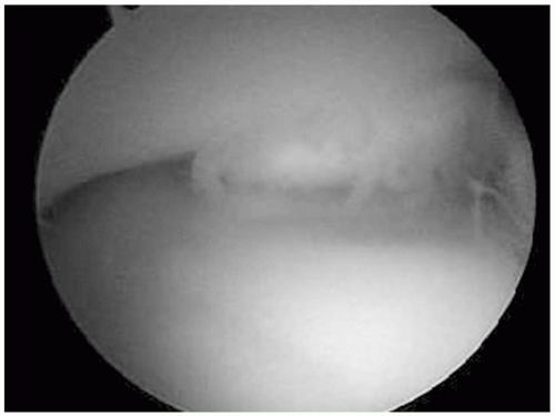
FIGURE 16-3 This arthroscopic view from the proximal anterior medial portal in the prone position demonstrates an anterior capitellar OCD lesion, located superiorly, with the radial head located inferiorly.


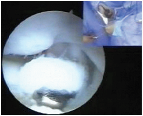
FIGURE 16-4 This arthroscopic view of a right elbow from the posterior central portal demonstrates a loose body in radial gutter, with a capitellar OCD lesion located superiorly and the radial head inferiorly.


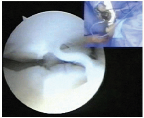
FIGURE 16-5 This arthroscopic view of a right elbow from the posterior central portal in the prone position demonstrates a lateral capitellar OCD lesion located superiorly and a large plica in the middle of the picture with the radial head located inferiorly.


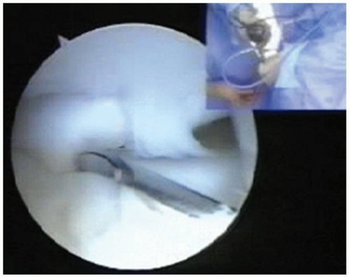
FIGURE 16-6 This arthroscopic view of a right elbow from the posterior central portal demonstrates spinal needle placement via the soft spot portal, to allow adequate instrumentation access to the lesion. The capitellar OCD lesion is located superiorly, with the radial head located inferiorly.


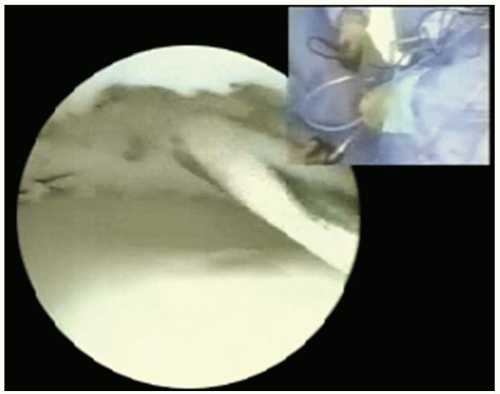
FIGURE 16-7 This arthroscopic view of a right elbow from the posterior central portal with a 70-degree arthroscope demonstrates antegrade drilling of an OCD lesion following debridement. The capitellar OCD lesion is located superiorly, with the radial head located inferiorly.


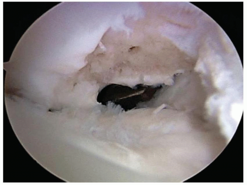
FIGURE 16-8 This arthroscopic view of a right elbow from the posterior central portal with a 70-degree arthroscope demonstrates an unconstrained OCD lesion following debridement and microfracture. The capitellar OCD lesion is located superiorly, with the radial head located inferiorly.

In the event that the lesion appears to have a large viable osteochondral fragment that is attached in situ, this can be slightly elevated to expose the undersurface and allow for debridement of the base. The fragment is then rotated into its anatomical position and stabilized with a percutaneous Kirschner wire. This can be converted to permanent fixation with a bioabsorbable implant, staples, a cancellous screw, or a Herbert-Whipple screw.

Cases of severe osteochondral defects may be amenable to osteochondral grafting from the proximal olecranon, knee or ribs, allograft, or synthetics. Placement of osteochondral graft plugs is best achieved through the distal soft spot portal, and elbow hyperflexion is often required for proper placement in the capitellar defect. The plugs are then contoured to match the remaining native capitellar surface (Figs. 16-9, 16-10 and 16-11). While synthetic grafting is approved for this use, this application remains experimental. In some cases, the lateral olecranon plug may include the proximal and lateral articular cartilage. This plug may be placed horizontally via a straight lateral distal portal with the lateral olecranon surface becoming the distal capitellar surface. In end-stage cases, the capitellum may be contoured using a shaver and a burr, excising the posterior capitellar spur and then resurfacing the bone with an allogeneic or synthetic patch.


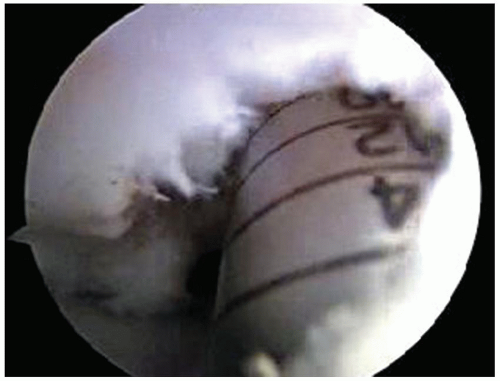
FIGURE 16-9 This arthroscopic view of a right elbow from the posterior central portal with a 70-degree arthroscope demonstrates preparation of an OCD lesion for placement of osteochondral plugs. The capitellar OCD lesion is located superiorly, with the radial head located inferiorly.
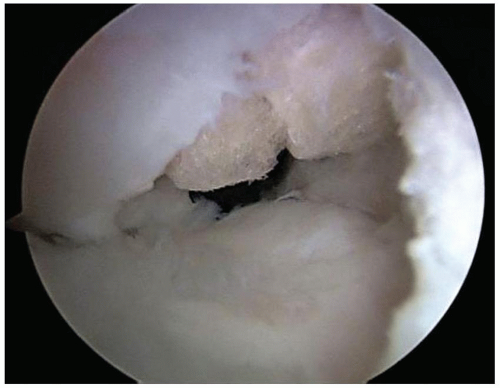


Get Clinical Tree app for offline access