CHAPTER 19 Osteochondritis Dissecans
INTRODUCTION
Osteochondral lesions may be the source of chronic elbow pain, swelling, and loss of motion in children or adolescents. The typical presentation is an adolescent gymnast or baseball pitcher.13,34 The dominant arm is usually involved, but may be bilateral in approximately 5% to 20%.34,41
OSTEOCHONDROSIS OF THE CAPITELLUM (PANNER’S DISEASE)
CLINICAL CHARACTERISTICS
Osteochondrosis of the capitellum is characterized by dull, aching pain in the elbow that usually is aggravated by motion or use, particularly by throwing or other violent motions. It usually occurs in boys and always during the period of active ossification of the capitellar epiphysis (ages 7 to 12, peak at 9 years). Panner’s disease is not seen before age 5 years. In addition to pain, loss of 5 to 20 degrees of elbow extension is common. Local swelling and tenderness over the lateral side of the elbow is not unusual.23 Symptoms resolve over time with few sequelae.
RADIOGRAPHIC CHARACTERISTICS
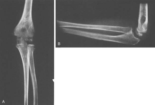
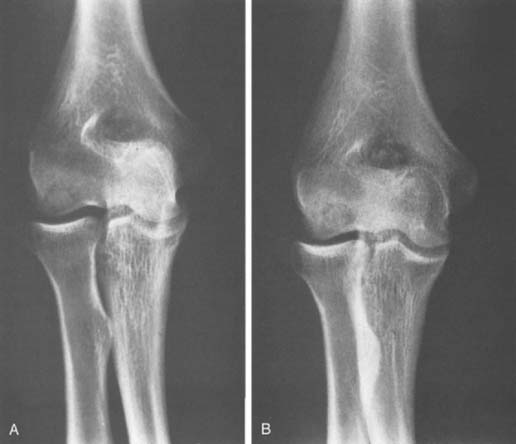
Osteochondrosis of the capitellum is a focal or localized avascular lesion of subchondral bone and its overlying articular cartilage.11 Radiographically, there is fragmentation of the capitellar epiphysis. The fragmentation is due to irregular patches of relative sclerosis alternating with areas of rarefaction (Fig. 19-1A and B). The outline of the epiphysis may be slightly irregular and smaller than that of the opposite normal capitellar epiphysis. Despite the radiographic fragmentation, osteochondral loose bodies do not form. As growth progresses, the capitellar epiphysis eventually assumes a normal appearance in size, contour, and internal architecture as clinical symptoms resolve. Residual deformity of the capitellum is rare.
Magnetic resonance imaging (MRI) findings include decreased signal intensity of the ossified epiphysis on T1-weighted images. Both plain films and MRI images of osteochondrosis of the capitellum are similar to findings in Legg-Calvé-Perthes disease of the hip.25 Deformity and collapse of the articular surface is less common in osteochondrosis of the capitellum than in Perthes’ disease of the hip.
OSTEOCHONDRITIS DISSECANS
INTRODUCTION
Osteochondritis is defined as an inflammation of both bone and cartilage. Osteochondritis dissecans is described as osteochondritis resulting in the splitting of pieces of cartilage into the joint (in Dorland’s Medical Dictionary). The term osteochondritis dissecans was given to this condition by Franz Konig in 1889, who described a knee condition that appeared to suggest a subchondral inflammatory process that dissected a fragment of cartilage from the femoral condyle, leading to formation of a loose body.20
Although no inflammatory process has ever been shown to produce such lesions, the name has remained. Osteochondritis dissecans of the capitellum is similar to osteochondritis dissecans in other joints, such as the knee. It involves localized avascular necrosis of subchondral bone and subsequent loss of structural support for the adjacent articular cartilage. Compared with the knee, however, osteochondritis of the capitellum is much less common. Only 6% of patients with osteochondritis dissecans have elbow involvement.41 Because osteochondritis dissecans occurs after the capitellum has almost completely ossified (early adolescence), it should not be confused with osteochondrosis of the capitellum, nor is it due to an “inflammation,” in spite of the definition of osteochondritis cited earlier.41 Osteochondrosis dissecans and osteonecrosis of the capitellum have been suggested as more appropriate terms, but are rarely used today.
From a practical standpoint, the localized area of osteochondritis, consisting of articular cartilage and underlying bone, either remains in situ and eventually heals or separates from the capitellum and becomes a loose body in the joint. Osteochondritis dissecans is one of several conditions that can cause “Little League elbow” in immature baseball players.38
CLINICAL CHARACTERISTICS
Elbow pain, the most common complaint, is usually dull, poorly localized, and aggravated by use, particularly by athletic endeavors that involve throwing or weight bearing on the upper extremity. Use aggravates the condition and rest relieves it. Lateral elbow pain occurs in approximately 79% to 90% of patients.18,41
A second complaint, limitation of elbow motion, particularly extension, affects about 90% of patients. It is often associated with an effusion, and results in 5 to 20 degrees loss of elbow extension.18,41 Limitation of elbow flexion, pronation, and supination of the forearm also occur, but these problems are less common.
RADIOGRAPHIC CHARACTERISTICS
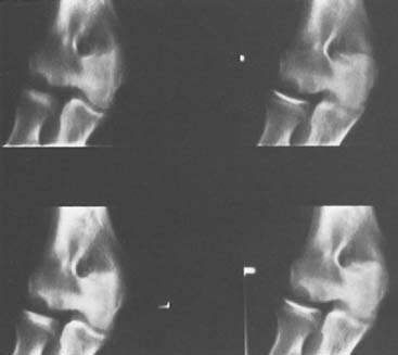
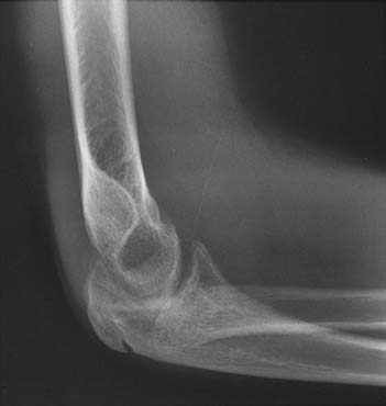
Anteroposterior and lateral radiographs of the elbow are useful and should be obtained in every case. Early in the disease, radiographic changes are most often confined to the capitellum.29 Rarefaction, irregular ossification, and a bony defect adjacent to the articular surface are frequent findings (Fig. 19-2). The crater of rarefaction in the capitellum usually has a sclerotic rim of subchondral bone adjacent to the articular surface.
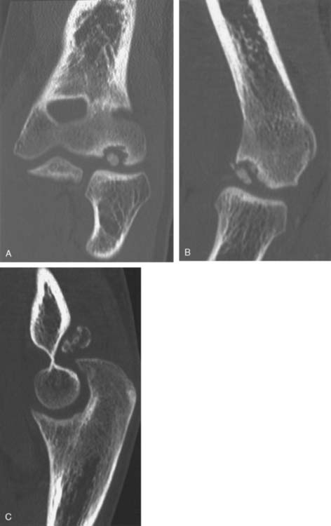
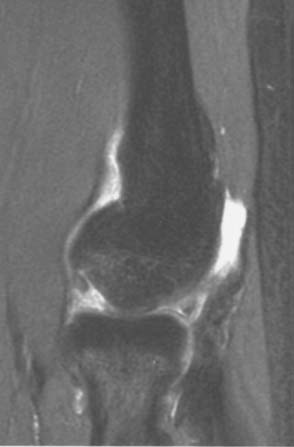
If the central fragment separates, one or more ossific loose bodies may be seen in the joint, and the articular surface of the capitellum may appear to be irregular or flattened, particularly on the lateral view. Only 30% of loose bodies can be seen on plain films.18 Computed tomography (CT) may be necessary to better define the bony anatomy (Fig. 19-3). MRI is the preferred imaging technique for assessing the extent of osteochondritis. MRI is also the best study to assess the integrity of the articular surface and the displacement of osteochondral fragments. Unstable lesions reveal fluid surrounding the osteochondral fragment on T2-weighted images (Fig. 19-4).8
Several other late radiographic changes are worth noting. As degenerative changes occur, the radial head enlarges. Klekamp described seven patients who had developmental dislocations of the radial head as a result of osteochondritis dissecans of the capitellum (Fig. 19-5).15 The cause-and-effect relationship is obscure. In a few instances, premature distal humeral physeal arrest is evident. Late in the course of disease, degenerative changes characterized by irregularity and incongruity of both the capitellar and radial head articular surfaces are evident; these changes are the most important late sequelae of this condition.
If sequestration does not occur, the central sclerotic fragment gradually becomes less distinctive, the surrounding area of rarefaction slowly ossifies, and the lesion heals without significant sequelae. This radiographic evidence of healing may take several years to occur and sometimes is not complete until adult life, long after pain, swelling, and limitation of motion have disappeared (Fig. 19-6A and B).
Stay updated, free articles. Join our Telegram channel

Full access? Get Clinical Tree


