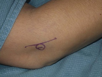
Chapter 49 Elbow pain is an extremely common problem in the general population, with lateral epicondylitis occurring in up to 4% of the general population. This condition can be exacerbated by athletic endeavors or activities involving heavy labor. Forceful activities and those with high forces combined with high repetition or awkward posture have been shown to increase the likelihood of development of lateral epicondylitis.1 Both in vivo and in vitro studies histologically illustrate an ongoing degenerative process followed by a reparative cycle.2 It is infrequent that a singular incident initiates a visit to the physician; rather, patients experience a chronic sequence of injury, pain, healing and resolution, followed by another inciting event. Prior investigations found that up to 26% of patients with this diagnosis have chronic symptoms and up to 40% have prolonged minor discomfort.3 In a review, Faro and Wolf found that “the vast majority of cases respond to conservative therapy…although it may take up to 18 months to attain full recovery.”4 Numerous studies have reported 75% to 90% cure rates with a sufficient trial of nonoperative therapy. Initial treatment of lateral epicondylitis begins with activity modification. Patients are instructed to avoid lifting with the wrist extended and to lift objects with the elbow in flexion as close to the body as possible with the hand in supination or neutral position to avoid applying stresses to the wrist extensor muscles. Patients who actively participate in racket sports should consider rackets with better shock absorption and consult with their instructor on optimal mechanics to minimize persistent problems. Conservative treatment also includes physical therapy focusing initially on stretching and progressive light eccentric strengthening of the forearm wrist extensors. Use of orthotics, such as nighttime wrist extension splinting, may be helpful for controlling pain.5 However, if nonoperative treatment for a period of 12 to 18 months fails, operative treatment is indicated for persistent pain and disability. Many techniques have been described for release of the extensor carpi radialis brevis (ECRB) that use open, percutaneous, and arthroscopic approaches. Recent research has shown no significant difference in outcomes between open and arthroscopic release.6 We prefer open excision of the pathologic portion of the ECRB tendon with subsequent stimulation of bleeding bone and repair of the defect, given the reproducibility of the postoperative results and relative straightforwardness of the procedure. History and Physical Examination Athletes typically have lateral elbow pain, frequently extending into the dorsal forearm, and decreased grip strength. Symptoms are exacerbated by activities involving wrist extension against gravity. The key finding during physical examination is localized tenderness at the origin of the ECRB. In addition, there is pain with wrist extension and long finger extension when the elbow is maintained in an extended position owing to the fact that the ECRB origin is at the base of the long finger metacarpal.7 The best candidate for operative intervention has had approximately 12 months of conservative treatment. This includes rest, nonsteroidal anti-inflammatory medication, bracing, and therapy.8 Other modalities such as extracorporeal shock wave therapy, botulinum toxin injection, corticosteroid injection, and platelet-rich plasma injection have been reported, but the evidence that these interventions are successful is limited. Other causes of lateral elbow pain such as radiocapitellar arthritis should be considered. Results after surgical intervention reveal that 40% to 97% of patients who undergo open ECRB release or debridement have decreased pain and improved function.4 Several surgical techniques have been described for the treatment of lateral epicondylitis. Surgical treatment ranges from resection of the epicondyle to percutaneous or open division of the common extensor origin to tendon-lengthening techniques. The most common technique involves identification and excision of the abnormal ECRB origin with creation of a vascular bone bed to promote healing.9 The authors prefer open excision of the pathologic portion of the ECRB tendon with stimulation of bleeding bone and repair of the defect for initial surgical management. Retrospective results have revealed high success rates, with 83% to 94% of patients experiencing pain relief.4 The specific steps of this procedure are outlined in Box 49-1. The skin incision is marked slightly anterior to the lateral epicondyle (Fig. 49-1) in a curvilinear pattern. Important landmarks to be outlined are the lateral epicondyle, radial head, and olecranon. Thick skin flaps are developed sharply down to the fascial layer. Typically, no significant cutaneous nerves are present in this area because it is a watershed region between the posterior antebrachial cutaneous nerve and the lateral antebrachial cutaneous nerve (Fig. 49-2).
Open Treatment of Lateral and Medial Epicondylitis
Lateral Epicondylitis
Preoperative Considerations
Key Physical Findings
Indications
Surgical Technique
Specific Steps
![]()
Stay updated, free articles. Join our Telegram channel

Full access? Get Clinical Tree


Open Treatment of Lateral and Medial Epicondylitis


