Chapter 84 Normal Knee Embryology and Development
The term embryology infers the study of embryos. Today, embryology generally refers to the entire period of prenatal development, however, and includes the study of embryos and fetuses. Although prenatal development is more rapid than postnatal development and results in striking changes, the developmental mechanisms of the two periods are the same. “Embryology provides a mechanism to help understand the causes of variations in human structure. It illuminates gross anatomy and explains how both normal relations and abnormalities develop.”20
Overview of Embryology
Prenatal development consists of four sequential stages:
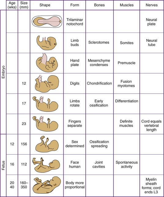
Figure 84-2 Prenatal development. This chart summarizes musculoskeletal development during embryonic and fetal life.
(From Staheli L: Growth. In Staheli L: Practice of pediatric orthopedics, Philadelphia, 2001, Lippincott Williams & Wilkins.)
Timing and Staging of Development
Gestational age based on the date of the mother’s last menstrual period overestimates the actual gestational age by more than 2 weeks. To estimate age more accurately, embryos are staged according to the method of Streeter.39 This system, a derivative of the Carnegie Embryonic Staging System, divides the embryonic period into 23 stages based on clearly defined details of either external form or the development of structures.26,27 The maturity of older embryos and fetuses is based on measurement of the crown-rump length.28
Normal Sequential Embryologic Development of the Knee*
Week 6
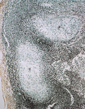
Figure 84-3 shows an embryo at 6 weeks old, Streeter stage 17. The cartilaginous anlagen of femur and tibia are separated by cells of uniform density, the future femorotibial joint. Early evidence of cavitation, a sign of beginning joint formation, in the otherwise homogeneous, uniform interzone is easily recognizable.
Week 7
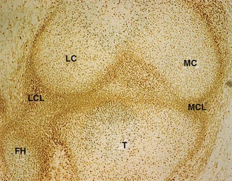
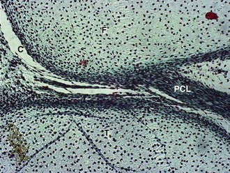
Figure 84-4 shows an embryo at 7 weeks old, Streeter stage 19. Already at this stage, the lateral femoral condyle and medial femoral condyle are well formed. The lateral collateral ligament spans from the femur to the fibular head, and the medial collateral ligament connects the femur to the tibia.
Week 8
Figure 84-5 shows an embryo at 8 weeks old. Not only is the posterior cruciate ligament seen, but also the multiple sites of beginning cavitation. Persistence of some of these intra-articular strands may lead to the development of plicae.
Week 10
Figure 84-6 shows a fetus 10 at weeks old. Between the medial femoral condyle and tibia, the medial meniscus can be seen. A small plica connects the midpart of the medial meniscus with the medial femoral condyle.
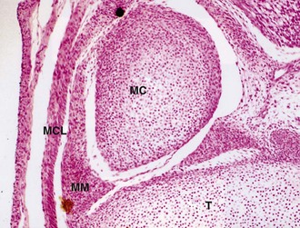
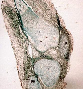
Figure 84-7 also shows a fetus at 10 weeks old. Not only the femur, tibia, and fibular head but also the anterior cruciate ligament are seen in the figure. At this stage, no vascular channels are present in the epiphyses.
Week 12.5
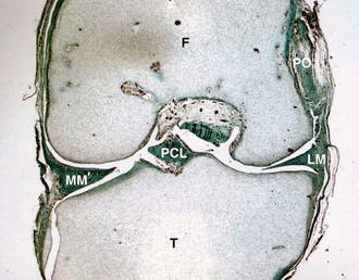
Figure 84-8 shows a fetus at 12.5 weeks old. A dense layer of cells covers the articular surfaces of the femur and tibia. Lateral to the lateral femoral condyle, the popliteus muscle is seen. Both menisci are well formed. Vessels are present at their periphery. Vascular channels are present in the femoral epiphysis. The posterior cruciate ligament inserts into the tibia.
Week 15.5
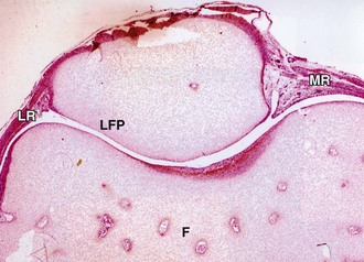
Figure 84-9 shows a fetus at 15.5 weeks old. The much longer lateral facet of the patella helps distinguish it from the medial facet. The lateral retinaculum is much denser than the medial retinaculum.
Week 16.5
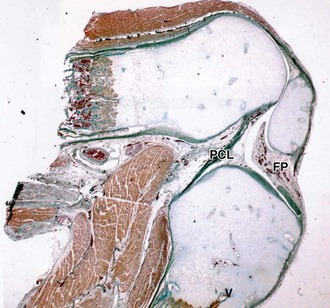
Figure 84-10 shows a fetus at 16.5 weeks old. The ossification processes that started in the diaphyses of the femur and tibia are progressing toward the metaphyses. At this stage, blood vessels still cross the growth plate. The suprapatellar bursa extends under the quadriceps muscle. The posterior cruciate ligament originates in the intercondylar fossa and infrapatellar fat pad.
Related posts:
 The Dislocated Knee
Treatment of Periprosthetic Fractures Around a Total Knee Arthroplasty
Imaging of Total Knee Arthroplasty
Repair and Reconstruction of the Medial Patellofemoral Ligament for Treatment of Lateral Patellar Dislocations: Surgical Techniques and Clinical Results
Cementless Total Knee Designs
Classification of Knee Ligament Injuries
The Dislocated Knee
Treatment of Periprosthetic Fractures Around a Total Knee Arthroplasty
Imaging of Total Knee Arthroplasty
Repair and Reconstruction of the Medial Patellofemoral Ligament for Treatment of Lateral Patellar Dislocations: Surgical Techniques and Clinical Results
Cementless Total Knee Designs
Classification of Knee Ligament Injuries
Stay updated, free articles. Join our Telegram channel

Full access? Get Clinical Tree


