This article provides an overview of the structure and function of the National Skeletal Muscle Research Center (NSMRC) at the University of California, San Diego, which is one of the 7 research centers of the Medical Rehabilitation Research Infrastructure Network, created to facilitate access for physicians to experts, technology, and resources from scientific fields related to medical rehabilitation. The 4 cores of the NSMRC are described as a resource for rehabilitation medicine practitioners to use for clinically relevant muscle research.
- 1.
Understand the organizational structure and function of the National Skeletal Muscle Research Center (NSMRC)
- 2.
Identify potential opportunities for collaboration with the NSMRC for clinicians interested in muscle research
Rehabilitation clinician-scientists often encounter a variety of primary and secondary diseases that are associated with skeletal muscle. Although muscular dystrophy, sarcopenia, and tendinitis are familiar skeletal muscle problems, diseases with cerebrovascular, coronary artery, and arthritic causes also affect skeletal muscle. Perhaps most importantly, most rehabilitation treatment strategies, regardless of the clinical entity, are directed toward or through skeletal muscle. Currently, scientific investigations in rehabilitation lack state-of-the-art, quantitative tools for measuring acute and chronic skeletal muscle changes. It is unreasonable to expect rehabilitation clinician-scientists to master the myriad of techniques necessary to study skeletal muscle or to put them in proper perspective when choosing methods. To address this methodological and knowledge void, The Medical Rehabilitation Research Infrastructure Network (MRRIN), funded by The National Institute of Child Health and Human Development (NICHD), through the National Center for Medical Rehabilitation Research (NCMRR), the National Institute for Neurologic Disorders and Stroke (NINDS), and the National Institute of Biomedical Imaging and Bioengineering (NIBIB), was created to facilitate access for physicians to experts, technology, and resources from scientific fields related to medical rehabilitation.
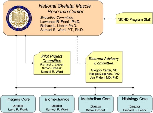
The MRRIN comprises 7 research centers, including the NSMRC of the University of California, San Diego (UCSD). The NSMRC consists of 5 core units that span 3 departmental boundaries and 2 schools within the university. The 5 cores are Muscle Histology, Muscle Biomechanics, Muscle Imaging, Muscle Metabolism, and Program Administration. The program is structured to foster cooperation and synergy across cores ( Fig. 1 ). This organization facilitates interaction among the program faculty and minimizes the overhead needed to accomplish rehabilitation research. Each of these cores is a strong research force at the UCSD and within the international research community. Thus, the strength of this infrastructure program is that it leverages existing strongholds of muscle research at UCSD and makes these resources readily available to the rehabilitation community within the United States. Another strength of the program is that it is based at UCSD, which is a major research University in the United States, and worldwide.
The NSMRC has been successful in equipping rehabilitation medicine professionals with the necessary tools to study muscle in the context of clinical problems and in attracting scientists to study skeletal muscle in a rehabilitation context. Since its inception in July 2005, the NSMRC has offered 6 symposia, and joined with such prestigious organizations as the Rehabilitation Institute of Chicago and the American Physical Therapy Association to sponsor 2 additional symposia. The NSMRC has also played a supporting role for several other NICHD-funded centers across the United States to provide muscle expertise or to train core directors. In addition, and most importantly, in the past 6 years, the program has funded $625,000 in pilot projects for 25 faculty members at 13 universities across the United States, resulting in many publications and National Institutes of Health (NIH) grant submissions. Although it was expected that career advancement and improved productivity would result from these pilot projects, some of the more subjective effects (prestige of independent funding, making scientific connections, learning and applying state-of-the-art methodologies) are almost equally important.
Core descriptions
Histology Core
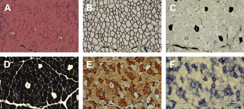
Skeletal muscle histology has a long and distinguished presence at the UCSD. Under the direction of Dr Richard Lieber and housed in the UCSD Department of Orthopaedic Surgery, skeletal muscle studies have been produced in large number since 1981. From the early studies describing skeletal muscle from normal and diseased tissues, to more recent studies that show more subtle changes that occur in skeletal muscle after injury from exercise or disease (as reported in Refs. ), the Histology Core has been at the forefront of the development of new methodologies and new approaches to studying skeletal muscle tissue ( Fig. 2 ). Many tools are available to the Histology Core. Traditional histologic methods such as paraffin embedding, frozen sectioning, and traditional histologic stains are available. However, the Histology Core is also able to perform immunohistochemical staining and correlate antibody stains with quantitative approaches and composition even on a single-cell basis. As an example of this technically demanding approach, the MRRPM NSMRC quantified the amount of various myosin heavy chains present in single cells of frog skeletal muscle fibers using correlative immunohistochemistry, quantitative Western blots, and reverse transcription/polymerase chain reaction (RT/PCR). There is thus virtually no protein structure or complex within skeletal muscle that cannot currently be studied by the Histology Core at the structural level, at least with the aid of the light microscope. In addition, the Histology Core has performed detailed quantitative analysis of skeletal muscle ultrastructure in exercised tissue and transgenic animal models. It has also combined forces with the Imaging Core and Biomechanics Core to develop methods for real-time imaging of single muscle cells during biomechanical loading. The Histology Core was used by 17 of the 25 projects supported by our infrastructure program from July 2005 to September 2011, making it the most heavily used core.
Biomechanics Core
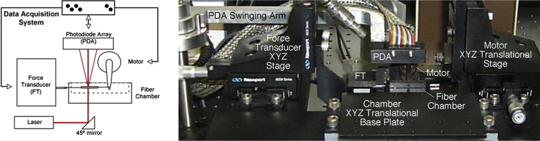
The muscle Biomechanics Core uses numerous approaches for biomechanical testing of muscles and muscle-related structures in a variety of settings. The Biomechanics Core is flexible and powerful in its ability to perform routine and advanced biomechanical studies. For example, standard facilities for performing laser diffraction of isolated skeletal muscles permit muscle architectural determination. Measurement of muscle architecture describes the most important single structural feature of a muscle that predicts function. To determine muscle architectural properties, fixed tissues are harvested, processed, and small bundles are isolated from the muscles. Muscle fiber bundles (consisting of 5–50 muscle fibers) are isolated and then sarcomere length (SL) of the isolated fiber bundles is determined by laser diffraction ( Fig. 3 ). This line of research has been extremely fruitful and was one of the main reasons that the Biomechanics Core received the 2003 Nicolas Andry Award from the Association for Bone and Joint Surgeons.
Related posts:
 Safety Concerns and Multidisciplinary Management of the Dysphagic Patient
Patient Safety in Cancer Rehabilitation
The Utility of Electromyography and Mechanomyography for Assessing Neuromuscular Function: A Noninvasive Approach
Skeletal Muscle Edema in Muscular Dystrophy: Clinical and Diagnostic Implications
Cardiac MRI in Muscular Dystrophy: An Overview and Future Directions
Neuromuscular Ultrasonography: Quantifying Muscle and Nerve Measurements
Safety Concerns and Multidisciplinary Management of the Dysphagic Patient
Patient Safety in Cancer Rehabilitation
The Utility of Electromyography and Mechanomyography for Assessing Neuromuscular Function: A Noninvasive Approach
Skeletal Muscle Edema in Muscular Dystrophy: Clinical and Diagnostic Implications
Cardiac MRI in Muscular Dystrophy: An Overview and Future Directions
Neuromuscular Ultrasonography: Quantifying Muscle and Nerve Measurements
Stay updated, free articles. Join our Telegram channel

Full access? Get Clinical Tree


