As more people survive burn injuries, there is an increasing focus on managing the complications of burn injuries with the ultimate goal of improving survivors’ quality of life. Musculoskeletal and neurologic sequelae are significant complications of burn injury. Electrical injury is a subcategory of burns with multiple musculoskeletal and neurologic complications. Knowledge of these complications helps clinicians provide optimal long-term care for burn survivors and enables survivors to attain maximal recovery.
Approximately one-half million people seek medical care for burn injuries each year, including 40,000 hospitalizations, 25,000 admissions to specialized burn centers, and 4000 deaths. With advances in acute management of burns over the past few decades more people are surviving severe burn injuries. As a result, providers are increasingly focused on the long-term management of burn survivors.
Burns are complex injuries that affect almost every organ system of the body and result in numerous long-term complications. Common complications of burn injuries include neurologic and musculoskeletal problems. These complications may develop a few weeks to years after injury, and significantly affects quality of life. Successful management of these complications directly impacts the burn survivor’s functional recovery.
The authors address burn-related neurologic and musculoskeletal complications in this article. In addition, electrical burn injuries are discussed because they are a subcategory of burns with significant neurologic and musculoskeletal complications.
Neurologic injuries
Clinically, neurologic complications are often underreported in the literature because the diagnosis is commonly delayed or missed entirely. The neurologic assessment is marred by the complexity of medical problems and impaired consciousness of patients who are critically ill. However, neurologic injuries cause serious debility and functional deficits that impact recovery. Both prevention and identification of neuropathies is an important aspect of burn rehabilitation. The neurologic complications of burn injuries are outlined in Table 1 .
| Complication | Comments |
|---|---|
| Mononeuropathy | Length of ICU stay, alcohol, electrical injury, age, and diabetes are risk factors. Specific positions predispose one to compression neuropathy. |
| Peripheral Polyneuropathy | Length of ICU stay and age are risk factors. |
| Mononeuritis Multiplex | Not well understood; lower-extremity injuries have better prognosis than upper-extremity injuries. |
| Pruritus | High incidence; managed with topical and oral medications |
Localized Neuropathies
Localized neuropathies are common after burn injuries with an incidence of 15% to 37%. Kowalske and colleagues examined 572 burn survivors and found that electrical injury, history of alcohol abuse, and length of intensive-care-unit (ICU) stay are significant risk factors for the development of mononeuropathies. Premorbid factors, such as elderly age and diabetes, are risk factors for the development of peripheral nerve compromise. In addition, prevention of compression neuropathy is an important tenet of rehabilitation. Bulky dressings can cause compression to superficial peripheral nerves, and improper and prolonged positioning can cause excessive stretch of nerves. Thus, proper positioning of patients as well as careful monitoring of wound care can mitigate neurologic complications. Clinical pearls of specific mononeuropathies and brachial plexopathy are reviewed in Table 2 .
| Neuropathy | Risk Factors |
|---|---|
| Brachial plexus | Shoulder abduction >90, external rotation Axilla/lateral chest wall grafting position |
| Ulnar nerve | Elbow flexion 90°, pronation, tourniquet paralysis |
| Radial nerve | At spiral groove: resting on side rails, hanging over edge of operating table, tourniquet paralysis At wrist: wrist restraints |
| Median nerve | Edema, prolonged or repeated wrist hyperextension, tourniquet paralysis |
| Peroneal nerve | Frog-leg position, lateral decubitus position, metal stirrups, leg straps, bulky dressings |
| Femoral nerve | Hematoma at femoral triangle, retroperitoneal bleed |
In general, poor positioning at the neck and shoulder leads to excessive stretch of the brachial plexus and places the plexus at risk for injury. Also, several bed and intraoperative positions commonly used in the treatment of burn injuries put the plexus at risk. Positioning of the shoulder during grafting of the axilla or lateral chest wall, with abduction of at least 90° and external rotation, places excessive stretch on the plexus. Patients are also placed in this position for alleviation of arm edema and prevention of axillary contractures. An alternate position that does not compromise the brachial plexus involves lying supine with the shoulder in 90° of abduction along with 30° of shoulder horizontal adduction.
In the upper extremity, the ulnar, median, and radial nerves are common sites for development of mononeuropathies. Positioning the elbow in flexion and pronation stretches the ulnar nerve and places it at risk for compression at the cubital tunnel. Median nerve injuries most commonly occur at the wrist. Local edema and prolonged or repeated hyperextension of the wrist compresses the nerve at the carpal tunnel. Clinicians should exercise caution when using wrist splints or an exercise program that includes hyperextension of the wrist. Radial nerve injuries most commonly result from compression at the spiral groove of the humerus. Therefore, arm positions that place the radial nerve at risk for injury include resting it on the bed siderails or hanging it over the edge of the operating table during interventional procedures. In addition, a pure sensory neuropathy results from compression of the superficial cutaneous branch of the radial nerve at the wrist, as with use of wrist restraints. Also, pneumatic tourniquets that are used in the operating room to establish a bloodless field may cause upper-extremity neuropathies. Improper inflation pressures can cause a direct pressure injury of the peripheral nerve at the cuff edge. The radial nerve is most vulnerable, but ulnar and median nerves are also at risk. Mononeuropathies of the upper extremities can also develop as a late complication of heterotopic bone formation. The elbow in particular is a common site of heterotopic ossification (HO) formation thus putting the ulnar nerve at risk.
In the lower extremities, the peroneal and femoral nerves are the most common mononeuropathies. The anatomic course of the peroneal nerve places it at risk with several common positions. Stretch injuries may occur with the frog-leg position, defined as externally rotated and flexed hip, flexed knee, and inverted foot. This position is used by patients with a tender medial thigh or perineal burns and when the bed is short for the patients’ height. Compression of the peroneal nerve at the fibular head is common, usually associated with the use of metal stirrups, leg straps, and the lateral decubitus position. Windowing of the dressing over the fibular head, used to relieve pressure, can also cause compression of the peroneal nerve. The femoral nerve is affected less often. Neuropathies develop in the femoral triangle typically because of compression by hematoma from venous or arterial blood draws. The discovery of a femoral nerve injury in patients on anticoagulation or with recent abdominal surgery raises suspicion of a retroperitoneal hemorrhage.
Peripheral Polyneuropathy
The pathophysiology of peripheral neuropathy in burns is complicated and thought to be caused by a combination of direct thermal injury on the nerves, circulating neurotoxins, and changes in distribution of fluid and electrolytes. These factors result from the body’s systemic response to the burn injury. After burn injury, a cascade of systemic physiologic processes ensues that affect the peripheral nervous system. There is a complex interplay of local and circulating mediators, including histamine, prostaglandins, thromboxane, kinins, serotonin, catecholemines, oxygen free radicals, platelet aggregation factors, angiotensin II, and vasopressin. Initially, there is vasoconstriction at the site of injury mediated by release of norepinephrine and serotonin. A few hours after injury vasoconstriction changes to vasodilation, causing increased capillary permeability and leakage of plasma into the extravascular space. Histamine is released and damaged cells swell. Fluid shifts result in increased extravascular edema and intravascular hypovolemia. Platelets and leukocytes aggregate, leading to thrombotic ischemia. In addition, inflammatory mediators are released. These effects lead to compromise of organ systems and, in particular, predispose the peripheral nerves to injury.
Generalized peripheral polyneuropathy is a common neurologic disorder in burn injury, with an incidence that ranges from 15% to 30%. Kowalske and colleagues found that age and length of intensive-care-unit stay are risk factors for developing polyneuropathies. Polyneuropathy is more commonly seen in those with greater than 20% total body surface area (TBSA) burns and electrical injuries. Electrophysiologic evidence of polyneuropathy is commonly seen within 1 week of severe burn injury. Clinically, patients may have symptoms of paresthesia and signs of mild to moderate weakness in the muscles of the distal extremities. On manual muscle testing, most patients eventually recover their strength, although they may complain of easy fatigability for years after the burn. Although critical-illness neuropathy is not explicitly documented in the burn literature, severely burned patients with prolonged intensive-care-unit stays, sepsis, and multiple organ failure are at risk for critical-illness neuropathy.
Mononeuritis Multiplex
Mononeuritis multiplex is an asymmetric sensory and motor peripheral neuropathy that involves 2 or more isolated peripheral nerves. The pathophysiology is not well understood but is thought to result from a combination of circulating neurotoxins, metabolic factors, and mechanical compression. Multiple mononeuropathy was documented in 7 of 121 subjects with greater than 40% TBSA burns in 1 study. In a separate study, mononeuritis multiplex was the most common diagnosis in subjects with burn injuries with a neuropathy. At 1 year after injury, lower-extremity nerve lesions demonstrated better functional recovery than upper-extremity nerve lesions.
Pruritus
The mechanism of pruritus is not well understood. Some investigators think it is related to axonal sprouting in the dermis and thereby classified here as a neurologic complication. Although not as devastating as some of the other neurologic manifestations of burn injuries, itch is nevertheless a significant complaint for many patients. The prevalence of pruritus is as high as 87% at 3 months and 70% at 1 year after injury. Predictors of pruritus include deep dermal injury, extent of burn, and early posttraumatic stress symptoms. Various treatment regimens have demonstrated a decrease in reported itch symptoms. However, a recent review examining pharmacologic and nonpharmacologic treatments of pruritus concluded that interventions lack strong empirical evidence. Treatments require better clinical studies to validate their use. Nonetheless, there exist multiple clinical treatment options. Nonpharmacological treatments, including colloidal oatmeal, pulsed dye laser, silicone gels, scar massage, and transcutaneous electrical nerve stimulation, demonstrate positive effects. Topical medications include histamine receptor antagonists and prudoxin, a tricyclic antidepressant with histamine blocking properties. There are reports of the use of topical anesthetics in the treatment of pruritis. Oral options also include selective histamine receptor antagonists and prudoxin. In addition, there is preliminary evidence for use of gabapentin and ondansetron for treatment of pruritis. For those with severe itching, often a combination of interventions is needed to control symptoms.
Musculoskeletal complications
Musculoskeletal complications are common after burn injuries. Prevention and early identification and treatment are the goals of care in the acute, subacute, and outpatient settings. Contractures are a major musculoskeletal complication of burn injury and are covered in their own article elsewhere in this issue. The authors address bone metabolism, osteophytes, heterotopic ossification, scoliosis and kyphosis, septic arthritis, and subluxations and dislocations in detail later ( Table 3 ).
| Complication | Comments |
|---|---|
| Changes in bone metabolism | Common in children; premature fusion of epiphyseal plate of long bones; low bone mineral density in large burns |
| Osteophytes | Most frequent skeletal change; most common at elbow |
| Heterotopic ossification | Most common at elbow Risk factors: burn size, ventilator support, ICU stay, prolonged wound closure, wound infection, and graft loss |
| Scoliosis and kyphosis | Developed in children with asymmetric burns and contractures |
| Septic arthritis | Caused by penetrating burns into a joint or hematogenous seeding; associated with joint dislocation, bone and joint destruction, and restriction of movement |
| Subluxations and dislocations | Most common in hand and feet because of contracture formation Prevention with splinting and range of motion |
Bone Metabolism
Delay in bone growth is a complication seen in the pediatric population following severe burn injury. Growth disturbances result from the premature fusion of the epiphyseal plate of affected long bones. Partial epiphyseal plate fusion may also occur, causing bone deviation and deformity. Bone-growth issues should be considered in growing children with burn scars that cross a joint and with joint contractures. In addition, case reports document that pressure garments for treatment of facial burns in children alter facial bone growth. Overbites may develop as a result of excessive pressure on the mandible. It is recommended to closely monitor facial development during and after pressure-garment use in children for development of normal dental and facial proportions. Pressure garments may need to be modified and changed frequently to avoid these complications.
Children with burns greater than 15% TBSA exhibit decreased bone mineral density. Investigators found decreased bone mineral density at 8 weeks after injury and the loss was sustained 5 years after injury. The mechanism for loss of bone mass is under investigation; however, recent research demonstrates causal roles for multiple factors, including increase in endogenous glucocorticoids, resorptive cytokines from the systemic inflammatory response, vitamin D deficiency, and disruption of calcium metabolism. Reduced bone density places children at risk for long bone fractures. Mayes and colleagues examined 104 burned children with greater than 40% TBSA and found a 5.8% incidence of fracture. Investigators have studied the use of recombinant human growth hormone without proven effect on bone formation. Recent studies have demonstrated improved bone mineral density with bisphosphonate therapy. Klein and colleagues performed a randomized controlled trial of 43 children with greater than 40% TBSA and examined the effects of acute administration (within 10 days of injury) of intravenous pamidronate. Subjects receiving pamidronate demonstrated higher whole body and lumbar spine bone mineral content at discharge, 6 months, and 2 years compared with controls.
Osteophytes
Evans and Smith reported that osteophytes are the most frequently observed skeletal alteration in adult patients with burn injuries. They are most often seen at the elbow and occur along the articular margins of the olecranon or coronoid process, and are thought to be caused by superimposed minor trauma to affected areas. Pain and nerve impingement can occur depending on the size and location of the osteophytes.
Heterotopic Ossification
Heterotopic ossification is the abnormal formation of bone in soft tissue. The incidence of HO is estimated at 1% to 2% of patients who are hospitalized with burn injuries. Clinically, only those with symptomatic joints, including impaired range of motion, joint pain, or other symptoms, require diagnostic evaluation. Therefore, reports in the literature reflect the incidence of clinically significant HO, not the true incidence. The etiology of HO is unknown. Investigators postulate that it is caused by the proliferation of primitive mesenchymal cells into osteogenic cells. Other factors thought to contribute to HO include hypercalcemia, prolonged immobilization, and remobilization after prolonged immobilization.
The elbow is the most frequent joint affected, comprising greater than 90% of cases in a 21-year review. Risk factors associated with the development of HO include burn size, ventilator support, intensive-care-unit stay, prolonged wound closure, wound infection, and graft loss.
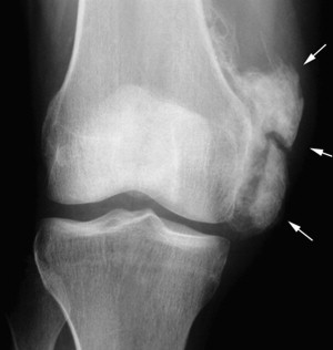
HO may occur as early as 5 weeks but usually develops approximately 3 months after injury. One of the earliest signs of heterotopic ossification is loss of joint range of motion. Other clinical findings may include swelling, erythema, pain, and peripheral nerve injury. Symptoms may precede radiologic findings. A bone scan is the most sensitive diagnostic imaging test and may demonstrate positive findings up to 3 weeks before positive radiographic findings. Three-phase bone scan is limited by its low specificity; one cannot differentiate HO from other traumatic, inflammatory, or degenerative processes. Plain radiographs demonstrate greater specificity than bone scan ( Fig. 1 ).
Treatment of HO begins with conservative measures, including positioning and range of motion to prevent worsening of joint motion. There are no studies examining HO prophylaxis in patients with burn injuries. However, there is evidence to support use of prophylaxis in other conditions and this data may help guide management of HO in the burn population. Nonsteroidal antiinflammatory drugs (NSAID) have proven efficacy for HO prophylaxis in patients with major hip surgery and spinal cord injury. A systematic survey of 13 randomized trials of NSAIDs used for HO prophylaxis in total hip arthroplasty showed a 57% reduction in HO in groups treated with NSAIDs. Bisphosphates are effective in reducing the incidence of HO in patients with spinal cord injuries. Subjects that received oral etidronate disodium for 12 weeks after injury exhibited lower rates of HO (6%) compared with controls (27%). Also, preoperative radiation of the affected hip reduces HO formation after total hip arthroplasty.
Surgical intervention is reserved for treatment of symptomatic HO. Heterotopic bone that causes nerve entrapment requires timely surgical intervention to avoid permanent nerve injury. It is common practice to wait until the bone is mature before surgical intervention for HO that results in impairments in upper-extremity and lower-extremity function, mobility, and activities of daily living. HO matures over 12 to 14 months and serial radiographs every few months are used to monitor for bone stabilization. Surgical excision of HO at the elbow results in improvement in range of motion. Tsionos and colleagues performed HO resection in 28 subjects and 35 elbows at a mean of 12 months after injury. At a mean follow-up of 21 months, flexion/extension improved from 22° preoperatively to 123° postoperatively. In a separate study of 8 children with elbow HO, all subjects demonstrated improved range of motion and were able to reach their face and perineum for functional tasks of feeding and toileting at 17 months after surgery.
Scoliosis and Kyphosis
Asymmetric burns of the trunk, hips, and shoulder girdle can cause patients to favor the affected side. In the growing child, the contracture of burn scars and resultant postural change can result in structural scoliosis. In a case series, 4 children who were scalded on the back as infants developed adolescent scoliosis. The deformities of all 4 cases were corrected surgically with good results. Similarly, childhood burns of the anterior neck, shoulders, and chest wall may produce a rounding of the shoulders and sunken chest. Likewise, burn-scar shortening and protective posturing can result in kyphosis. Both scoliosis and kyphosis are amenable to bracing and surgical interventions. An orthopedic surgeon is recommended to follow such patients.
Septic Arthritis
Septic arthritis is challenging to diagnose in patients who are severely burned. The characteristic signs and symptoms are often absent or masked by the overlying burn wound. Joint pain, swelling, color change, and tenderness are common symptoms at the site of burn injury or grafting and therefore are difficult to distinguish from septic arthritis.
The 2 major causes of a septic joint are penetrating burns into a joint and hematogenous seeding from bacteremia. Patients with burn injuries are at risk for infection because of their impaired immune system and concurrent illness. Septic arthritis may cause gross dislocation because of capsular laxity or cartilage and bone destruction, or result in severe restriction of movement or ankylosis. It occurs most frequently in the joints of the hands, hips, knees, and wrists.
Subluxations and Dislocations
Joint subluxation of the hands and feet are common after burn injury. Burns of the dorsal surface may contract resulting in joint hyperextension. Prolonged hyperextension places the joint at risk for subluxation. This result is most common at the metacarpophalangeal (MCP) and metatarsophalangeal (MTP) joints. Ulnar neuropathy places patients at additional risk for subluxation of the fourth and fifth digits. For dorsal hand burns, prevention of subluxation is achieved with a combination of splinting and range-of-motion exercises. A dorsal hand burn splint places the MCP joints in 60° to 90°of flexion and the distal and proximal interphalangeal joints in full extension. Similarly, the MTP joints may subluxate after contracture of healed wounds, especially in children. Application of surgical high-top shoes with a metatarsal bar helps prevent toe deformities.
Posterior hip dislocation is a problem in children. Hips maintained in an adducted and flexed position are at risk for dislocation. Anterior shoulder dislocations occur in positions of abduction and extension. Shoulder dislocations may result from positioning in the operating room.
Musculoskeletal complications
Musculoskeletal complications are common after burn injuries. Prevention and early identification and treatment are the goals of care in the acute, subacute, and outpatient settings. Contractures are a major musculoskeletal complication of burn injury and are covered in their own article elsewhere in this issue. The authors address bone metabolism, osteophytes, heterotopic ossification, scoliosis and kyphosis, septic arthritis, and subluxations and dislocations in detail later ( Table 3 ).
| Complication | Comments |
|---|---|
| Changes in bone metabolism | Common in children; premature fusion of epiphyseal plate of long bones; low bone mineral density in large burns |
| Osteophytes | Most frequent skeletal change; most common at elbow |
| Heterotopic ossification | Most common at elbow Risk factors: burn size, ventilator support, ICU stay, prolonged wound closure, wound infection, and graft loss |
| Scoliosis and kyphosis | Developed in children with asymmetric burns and contractures |
| Septic arthritis | Caused by penetrating burns into a joint or hematogenous seeding; associated with joint dislocation, bone and joint destruction, and restriction of movement |
| Subluxations and dislocations | Most common in hand and feet because of contracture formation Prevention with splinting and range of motion |
Related posts:

Stay updated, free articles. Join our Telegram channel

Full access? Get Clinical Tree


