The evolving shift in focus of burn care and research toward optimizing the long-term outcomes of persons with burn injuries has certainly increased the emphasis on burn reconstruction. There are an increasing number of persons surviving extensive injury who may have long-term reconstructive needs. Burn reconstruction, just as acute burn care, requires a coordinated team approach from initial consultation through recovery and rehabilitation. Clearly, in the future, one can expect evolution in surgical techniques and technologies that can improve the function and appearance of persons with burn injury.
The tremendous advances in survival following burn injury over the past decades have shifted emphasis in care of the burn patient form survival alone to optimization of functional and psychosocial outcomes. Accordingly, reconstructive surgery plays a rightful prominent role in modern burn care. Burn reconstruction refers to the numerous procedures performed on persons with burn injuries to optimize their functional outcomes and appearance. It should be emphasized that these procedures should always be considered “reconstructive” rather than “cosmetic” because they are performed to restore what has been lost or damaged as a result of injury rather than to improve uninjured tissue for purely aesthetic reasons.
Reconstructive surgery—just as acute burn care—requires a multidisciplinary team approach. Psychological preparedness for surgery, adequate nutrition, postoperative splinting, and range of motion programs require the participation of the burn team.
Timing of reconstructive surgery
Reconstructive procedures can occur several weeks to several years—even decades—following injury. There are several important considerations to selecting the appropriate time for surgery related to both the patient’s physical and psychological condition. Because most burn procedures deal with issues related to scars and because the scar maturation process typically requires 12 to 18 months for completion, most procedures are not performed for at least a year following injury, as the need for procedures or the extent of procedure required may decrease over time. However, there are several important instances where surgery should be performed earlier such as in the case of exposed vital structures, severe eyelid ectropion, or unstable wounds.
There are several critical perquisites that must be met before undertaking reconstructive procedures. First and foremost is the psychological preparedness of the patient. Anatomic defects alone are not sufficient critieria to proceed with surgery. Patients need to be prepared to undergo procedures—including being prepared to return to the hospital and miss periods of work and school. Many patients will rightfully have anxiety about undergoing additional operations and have trepidation about returning to the hospital ward where they may have previously spent weeks to months. In some cases, for patients who will require many procedures over time, selecting to do a smaller procedure that does not require hospital admission and has a predictable outcome may be a reasonable first reconstruction to perform.
Patients must also have realistic expectations as to what can be achieved with each procedure. Although many patients will come to the burn reconstruction clinic with the hope that scars can be simply erased and their preinjury appearance restored, the unfortunate reality is that this will likely not be possible. Therefore, a frank discussion with the patient and his or her family is required as to what can reasonably be accomplished needs to occur. In addition, the likely need for multiple operations spread out over months to years also needs to be addressed. Often at the first visit to the burn reconstruction clinic we, along with the patient, will formulate a list of problems and potential surgeries that can be performed over time and work with the patient to prioritize these procedures. This list remains in the front of the patient chart and can be revisited and revised as needed over time. When possible, procedures should be grouped to minimize the overall number of surgeries. However, procedures that require immediate postoperative mobilization, such as joint capsulotomies and capsulectomies, should not be grouped with procedures that require prolonged postoperative immobilization like joint arthrodesis or contracture release with skin graft. There are also instances where there is no good or safe reconstructive option and this too must be carefully and thoughtfully explained to the patient. However, it is critical to still provide some hope when possible that new procedures or technologies may be developed in the future.
Special consideration needs to be made in the case of children and adolescents. It is critical that children be included in the decision-making process for surgery. Often parents will bring their children to clinic and the child may not be interested or ready for surgery. We make every effort to emphasize to parents that their child must “buy-in” to having procedures done because they are the ones having to undergo the procedure and their cooperation with postprocedural splinting and/or range of motion is also critical to the success of the procedure.
Reconstructive needs
There are a number of common deformities that occur following burn injury that may be amenable to reconstructive surgery.
Scars and Contracture
Scarring remains the number one source of discomfort and misery for persons with burn injury ( Fig. 1 ). Despite all of the advances in burn care over the past few decades and the high incidence of scarring, reported to be as high as 50% following burn injury, there is still a limited understanding as to the risk factors and pathophysiology of scar formation. Accordingly, there exist few effective strategies to prevent scar formation and few proven strategies for treating them. Known risk factors for scarring include delay in wound closure, infection, and race (patients with pigmented skin are believed to be at higher risk for scar formation). A number of nonsurgical therapeutic options have been described, including massage, pressure, silicone, vitamin E, and steroids. Steroid injection and topical silicone can be used on scars during the maturation process to alleviate symptoms of pain and itch and potentially reduce scar size; however, scar injection is difficult in children and may not be practical for large areas of scar. More recently, the use of lasers to improve scar appearance has been described. Parrett and Donelan have reported using the pulsed dye laser to reduce scar rubor in the early postinjury period. Whether or not treatment early on with a laser can affect ultimate scar burden remains to be seen with more experience.
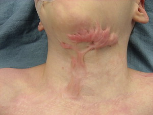
Wound contraction is a natural process of shrinking that occurs in all healing tissues, whether or not they have been treated with a skin graft. Contracture refers to deformities that occur as a result of wound contraction across a joint or other mobile surface. Burn contractures occur most commonly in the hands, axilla, neck, and face. Limited contractures can often be overcome with aggressive range of motion exercises and splint immobilization; however, more extensive contractures will require surgical release. It is important to note that following surgical release, aggressive range of motion and periods of splint immobilization may be required to minimize contracture recurrence.
Pigment and Hair Loss
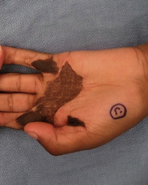
Problems of uneven pigment—both hyperpigmentation and hypopigmentation—are common complaints in persons with burn injury. Pigment problems can develop in areas that have healed spontaneously, areas that have undergone grafting, and in donor sites ( Fig. 2 ). There are limited treatment options for pigment problems. Whitening agents such as hydroquinones have been used with some success for areas of hyperpigmentation and there have been early reports of success with dermabrading areas of hyper- or hypopigmentation and regrafting with epithelial cells obtained from the same anatomic area. However, additional clinical experience is needed to verify the success of this approach.
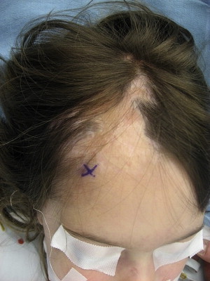
Hair loss can occur in areas that were burned and grafted or in areas of deep burn that healed without grafting ( Fig. 3 ). Small areas of hair loss can be addressed by excision of the hairless areas and larger areas can be managed with tissue expansion, as detailed later in this article. The use of hair micrografts for eyebrows and moustache areas have also been used with success. Inappropriate hair growth in thick (ie, full-thickness) grafts can also occur. These areas can be treated with common depilatory techniques, including laser treatment or manual depilation.
Pain and Itch
Pain and itch remain common complaints even years after burn injury. Although wounds may be all closed and scars well matured, patients will still complain of persistent pain and itch. If pain and itch occur in a discrete area of scar then surgical excision of the scar may provide significant relief. Similarly, pain associated with a contracture deformity may also resolve with surgical correction. However, pain alone should not be an indication for surgery. In the absence of a visible deformity that can be corrected surgically, undertaking a procedure to relieve pain may prove fruitless.
Reconstructive needs
There are a number of common deformities that occur following burn injury that may be amenable to reconstructive surgery.
Scars and Contracture
Scarring remains the number one source of discomfort and misery for persons with burn injury ( Fig. 1 ). Despite all of the advances in burn care over the past few decades and the high incidence of scarring, reported to be as high as 50% following burn injury, there is still a limited understanding as to the risk factors and pathophysiology of scar formation. Accordingly, there exist few effective strategies to prevent scar formation and few proven strategies for treating them. Known risk factors for scarring include delay in wound closure, infection, and race (patients with pigmented skin are believed to be at higher risk for scar formation). A number of nonsurgical therapeutic options have been described, including massage, pressure, silicone, vitamin E, and steroids. Steroid injection and topical silicone can be used on scars during the maturation process to alleviate symptoms of pain and itch and potentially reduce scar size; however, scar injection is difficult in children and may not be practical for large areas of scar. More recently, the use of lasers to improve scar appearance has been described. Parrett and Donelan have reported using the pulsed dye laser to reduce scar rubor in the early postinjury period. Whether or not treatment early on with a laser can affect ultimate scar burden remains to be seen with more experience.
Wound contraction is a natural process of shrinking that occurs in all healing tissues, whether or not they have been treated with a skin graft. Contracture refers to deformities that occur as a result of wound contraction across a joint or other mobile surface. Burn contractures occur most commonly in the hands, axilla, neck, and face. Limited contractures can often be overcome with aggressive range of motion exercises and splint immobilization; however, more extensive contractures will require surgical release. It is important to note that following surgical release, aggressive range of motion and periods of splint immobilization may be required to minimize contracture recurrence.
Pigment and Hair Loss
Problems of uneven pigment—both hyperpigmentation and hypopigmentation—are common complaints in persons with burn injury. Pigment problems can develop in areas that have healed spontaneously, areas that have undergone grafting, and in donor sites ( Fig. 2 ). There are limited treatment options for pigment problems. Whitening agents such as hydroquinones have been used with some success for areas of hyperpigmentation and there have been early reports of success with dermabrading areas of hyper- or hypopigmentation and regrafting with epithelial cells obtained from the same anatomic area. However, additional clinical experience is needed to verify the success of this approach.
Hair loss can occur in areas that were burned and grafted or in areas of deep burn that healed without grafting ( Fig. 3 ). Small areas of hair loss can be addressed by excision of the hairless areas and larger areas can be managed with tissue expansion, as detailed later in this article. The use of hair micrografts for eyebrows and moustache areas have also been used with success. Inappropriate hair growth in thick (ie, full-thickness) grafts can also occur. These areas can be treated with common depilatory techniques, including laser treatment or manual depilation.
Pain and Itch
Pain and itch remain common complaints even years after burn injury. Although wounds may be all closed and scars well matured, patients will still complain of persistent pain and itch. If pain and itch occur in a discrete area of scar then surgical excision of the scar may provide significant relief. Similarly, pain associated with a contracture deformity may also resolve with surgical correction. However, pain alone should not be an indication for surgery. In the absence of a visible deformity that can be corrected surgically, undertaking a procedure to relieve pain may prove fruitless.
Overview of techniques for reconstructive surgery
There are a number of common techniques used in burn reconstructive surgeries. A brief description of the most common techniques used is provided in the following sections.
Scar Release and Excision
Scar contracture release is usually achieved by incising the scar band at its point of maximal tension. Given that contracture bands typically exist in a larger area of scar, releasing incisions are often extended to tissue beyond the scar band itself. Full release requires that incision be carried completely through the scar tissue until healthy tissue is reached. In some cases, release of the subcutaneous tissue and muscle fascia may also be required to achieve full release. This is often the case in the neck and axilla. Long-standing deformities of the digits may also require dividing the scar around the tendons and joint capsules and the surgeon must be aware of the potential need to divide these underlying structures and challenges in covering the subsequent defect, as skin grafting is not possible on exposed tendon, bone, and joint.
For broad areas of scar, total scar excision is necessary and practical. These procedures are relatively straightforward and have predictable results. However, although they may be relatively simple from a technical standpoint, they can provide tremendous relief to patients suffering from pain and itch from a hypertrophic scar. There are 2 general approaches to scar excision—extralesional and intralesional. Extralesional excision refers to excision of the entirety of the scar and suturing together healthy, unscarred tissue ( Fig. 4 ). In contrast, intralesional excision refers to excision of most of the scar but a rim of scar is left behind and sutured together. The purported benefit of intralesional excision is that there will be no new tissue injury—incision is made through previously scarred tissue only and therefore the process of scar formation has already occurred in this tissue and is unlikely to recur. The relative risks and benefits of each approach should be discussed with the patient before surgery. Many scars are too large to be removed in one setting and often require several “serial” excisions. We typically wait 8 to 12 months between serial excisions so the tissue can sufficiently heal and soften so that it can be optimally mobilized again to achieve closure.


