Chapter 31 Nerve Entrapment Around the Elbow
Introduction
The elbow is a common site of nerve entrapment syndromes. The reasons for this are multifactorial, and this chapter will identify the more common causes and their management, with an emphasis on our personal approach.
The three major nerves of the upper limb (ulnar, median and radial) pass from the arm into the forearm in close proximity to the elbow joint. The ulnar nerve passes between the two heads of flexor carpi ulnaris (FCU) with the median nerve passing between the two heads of pronator teres. The radial nerve or, more accurately, its posterior interosseous motor branch, passes between the two heads of the supinator muscle. Each nerve and its branches can be compressed, giving rise to well-described nerve entrapment syndromes.
Background/aetiology
The nerves that cross the elbow are either purely sensory, purely motor or contain mixed sensory and motor fibres. When nerve entrapment occurs the classical presentation involves loss of sensation and motor power. The modalities of sensation travel in the larger diameter fibres and are often the first and most severely affected by compression. These include light touch and fine discrimination. Smaller diameter fibres that convey pain are well preserved until severe prolonged compression occurs. Loss of motor power is identified in the specific muscle groups that are supplied by the compressed nerve. Macroscopic loss of power can be noticed within the large muscle groups, including the forearm flexors and extensors. These may manifest themselves as finger drop, loss of grip strength or generalized weakness. Weakness in the ulnar innervated muscles of the hand may be detected before wasting is seen if a careful clinical history is obtained. Patients often describe progressive clumsiness in the hand with loss of the normal fine motor control. This is followed by wasting of the small muscles of the hand that is easily visualized due to the lack of fat in this region or by comparison of asymmetry with the opposite limb.
Clinical examination of the hand may also reveal trophic changes. These include healing burns, scalds and lacerations that the patient may not have noticed until the damage had been done. A thorough history and clinical examination should enable an appropriate differential diagnosis of the possible sites of compression. These include the anterior horn cell, the spinal canal, the intervertebral foramen and the thoracic outlet together with the elbow, wrist and hand. In addition to a careful assessment of the nerves around the elbow, physical examination should include a general neurological assessment with examination of the legs for any long tract signs that may suggest a lesion in the spinal cord or canal.
The clinician should also be aware that occasionally a ‘double-crush’ phenomenon may occur. This was first described by Upton and McComas1 in 1973 who stated that ‘neural function was impaired because single axons, having been compressed in one region, become especially susceptible to damage at another site’. When this occurs in the upper limb the peripheral nerves become hypersensitized by proximal compression in the neck and are more susceptible to an otherwise well-tolerated level of compression.
Clinical Pearl 31.1
In the assessment of nerve entrapment around the elbow, clinical examination should include the sensory and motor supply of the ulnar, median and radial nerves together with a careful examination of the cervical spine and lower limbs. The clinician should also be aware of the possibility of a ‘double crush’.
The causes of compression can vary widely and are given below.
The anatomical structures around the elbow and, in particular, the various fibro-osseous tunnels and fibrous arches can cause rigid borders against which nerves may be compressed. The large range of elbow flexion will produce longitudinal traction on the nerves lying within the extensor compartment whilst compressive forces are applied to the nerves on the flexor surface. The ulnar nerve for example is subjected to longitudinal traction during terminal flexion. In addition it has been reported that 5.1 mm of ulnar nerve excursion is needed for elbow motion from 10° to 90°.2 This alone may compromise nerve function, but when combined with a second local insult such as a fibrous band or impinging osteophyte there is an increased likelihood of nerve irritation.
Congenital abnormalities can also produce nerve entrapment. The median and very occasionally the ulnar nerve may be compromised by the ligament of Struthers. This arises from a supracondylar spur on the medial border of the distal humeral shaft and extends obliquely to the medial epicondyle (Fig. 31.1).

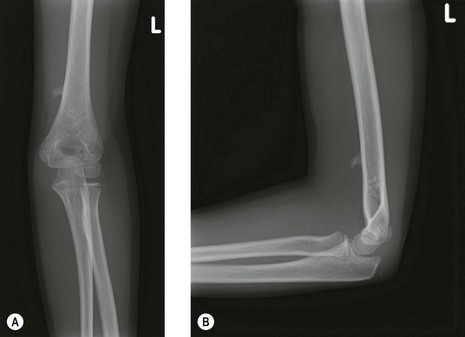
Figure 31.1 (A) Supracondylar process: anteroposterior view. (B) Supracondylar process: lateral view.
Inflammatory processes, particularly rheumatoid arthritis, have been implicated in nerve entrapment at the elbow. Posterior interosseous nerve palsy secondary to radiocapitellar synovitis, can give rise to a lag in finger extension and lead to a misdiagnosis of tendon rupture at the wrist. Careful clinical assessment will distinguish the palsy from a tendon rupture or subluxed extensor tendons in the metacarpophalangeal joint gutters.
Transient ulnar nerve palsy may occur from a direct blow to the nerve in its relatively unprotected medial epicondylar groove. More serious is tardy ulnar nerve palsy that is usually the result of excessive cubitus valgus deformity. This occurs most frequently after non-operatively treated displaced lateral humeral condylar fractures of childhood.
A number of metabolic conditions are well-recognized causes of peripheral nerve entrapment syndromes. These include pregnancy, thyroid disease, diabetes and sarcoidosis. The symptoms of nerve compression may resolve once the underlying medical cause is treated although this is not always the case.
Cubital tunnel syndrome (ulnar nerve)
Most patients who present with cubital tunnel syndrome have no identifiable aetiology for their compression although in a minority a cause can be recognized, and these include:
Background and aetiology
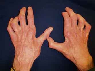
The ulnar nerve at the level of the elbow is a large mixed motor and sensory nerve. It provides sensation to the ulnar one and a half digits of the hand, the volar and dorsal aspect of the hand and the medial aspect of the forearm. It is the motor supply to FCU, flexor digitorum profundus (FDP), palmaris brevis, adductor pollicis, the deep head of flexor pollicis brevis, seven interossei, three hypothenar muscles and the lumbricals to the little and ring fingers. With severe ulnar nerve compression at the elbow, the commonest site of muscle wasting, is the first dorsal interosseous muscle (Fig. 31.3).
The intraneural anatomy of the ulnar nerve at the elbow is organized into a layered formation of fibres.3 Sunderland showed that the sensory supply to the hand was present in the most superficial layer beneath which was found the innovation of the intrinsic muscles. The motor branches to the long flexor tendons were present in the deepest portion of the nerve. This patterning explains the early onset of sensory symptoms in the hand and why weakness of the long flexor tendons occurs at a much later stage with more significant compression.
The ulnar nerve blood supply is segmental and is a major concern during anterior transposition when the nerve is solely dependent on its intraneural supply. Prevel et al4 in a study on the extrinsic blood supply showed that the ulnar nerve receives two constant major pedicels from the superior ulnar collateral artery proximally and the posterior ulnar recurrent artery distally. In a cadaveric study they demonstrated by measuring total vessel length and distance to the medial epicondyle that the extrinsic vascular supply could be preserved during anterior transposition of the ulnar nerve, even after the nerve had been extensively mobilized. Simple decompression, however, has the advantage of leaving the nerve in situ with its surrounding vascular supply.
The ulnar nerve can be compressed at four main sites around the elbow:
Presentation, investigations and treatment options
Presentation
Patients present with an insidious onset of altered sensation in the ulnar nerve distribution. The sensory loss on the dorsum of the hand helps differentiate compression of the ulnar nerve at the elbow from compression within Guyon’s canal at the wrist. Compression in Guyon’s canal spares sensory changes on the dorsum of the hand due to the fact that the dorsal sensory branch to the back of the hand arises proximal to the wrist.
Initial sensory symptoms are often reported as a ‘cotton wool’ feeling. Pins and needles occur in the same distribution and may lead to a gradual progressive loss of sensation. Symptoms are usually worse at night but are less classical than those that occur with carpal tunnel syndrome. The nocturnal symptoms occur in our opinion due to the loss of the muscle pumps at night and pooling of interstitial fluid in the peripheries.
Patients may also report motor symptoms and these should be taken seriously as muscle wasting can occur quickly. An early warning of impending motor loss is the feeling of clumsiness in the hand. Functional activities of daily living with the affected hand such as hair drying, brushing hair, driving and telephone use are all aggravated by elbow flexion.
In the early stages of cubital tunnel syndrome there may be few if any physical signs. Inspection may reveal excessive cubitus valgus. The normal elbow has approximately 7° of physiological valgus although this varies between individuals. The key is an asymmetrical valgus deformity. Careful questioning may identify a childhood fracture. Inspection may also reveal a deformed elbow joint consistent with osteoarthritis. Muscle wasting may be observed in the ulnar flexor forearm and in the hand. Wasting of the first dorsal interosseous muscle is very easily identified when present. The hand may take on a guttered appearance with intermetacarpal wasting of the interossei.
Palpation of the elbow may reveal ulnar nerve tenderness in the cubital tunnel, coexisting medial epicondylitis or an ulnar nerve which subluxes anteriorly on flexion. Tinel’s test may be positive anywhere along the nerve and in our experience it is more sensitive than when performed at the wrist for carpal tunnel syndrome. The test involves gently tapping along the course of the nerve from distal to proximal. An unpleasant sensation is felt at the site of entrapment.
Froment’s sign is also useful in the assessment of ulnar nerve function. It involves placing a piece of paper between the patient’s adducted thumbs and index fingers of both hands. The patient is then asked to resist extraction of the paper by the examiner. With normal power, the first dorsal interosseous and adductor muscles will prevent removal of the paper from between the digits. However, with ulnar nerve palsy these muscles are weak and the patient subconsciously recruits the flexor pollicis longus (FPL) tendon (anterior interosseous nerve supply). The thumb interphalangeal joint flexes to pinch the paper and prevent its extraction. Additional evidence of ulnar nerve dysfunction is the patient’s inability to cross their fingers symmetrically.
Our favoured provocation manoeuvre is the flexion compression test. In this test the patient is asked to fully flex the elbow while the examiner applies digital pressure over the cubital tunnel. A positive test is associated with the development of ulnar nerve symptoms within 30 seconds of applying digital pressure.
Investigations
Patients suspected of bony abnormalities, including cubitus valgus or osteoarthritis, should undergo anteroposterior and lateral plain radiographs centred on the elbow. In addition a cubital tunnel view is useful to visualize the medial epicondylar groove. This comprises a posteroanterior radiograph taken with the elbow in full flexion. A plain anteroposterior radiograph in full elbow extension will demonstrate valgus alignment. If this is greater than 7° a simple ulnar nerve decompression may be unsuccessful in relieving the patient’s symptoms and an anterior transposition is the preferred option.
Electro-physiological investigations are of value in clinically equivocal scenarios or where the site of the compression is in doubt or in the setting of a possible polyneuropathy or double crush syndrome.
An ultrasound scan may be a useful test to visualize the ulnar nerve in cases of suspected subluxation in the larger obese patient. It will also reveal the lack of nerve excursion and movement on flexion and will show sites of tethering and compression. In addition it may indicate neural swelling proximal to the site of compression.5
Treatment options
Non-operative treatment
The non-surgical management of ulnar neuropathy should be restricted to mild to moderate entrapment.9 Simple analgesics and non-steroidal antiinflammatory drugs may be useful for some patients. Others find splints beneficial although rigid devices are often poorly tolerated by both the patient and their bed-time partner. As a short-term measure we advise the patient to make a hole at the far end of a pillowcase. The hand can be passed through the pillowcase, alongside the pillow exiting on its far side through a small window that has been cut in the seam. During rest at night, flexion of the elbow, which often accompanies patients who sleep in the fetal position, is prevented. The elbow is unable to flex due to the bulky pillow. Patients often find this useful and more comfortable than other types of splint. In our experience, however, spontaneous resolution of symptoms is unlikely if the nerve entrapment syndrome is secondary to osteoarthritis, if there is a significant bony deformity such as cubitus valgus; or, if the compression appears to be severe, with significant motor wasting or sensory loss.
Surgical technique and rehabilitation
Although cubital tunnel surgery is usually performed under general anaesthesia as a day case, it can be performed under local anaesthesia in patients with comorbidities that prevent the safe use of a general anaesthetic. It is unwise, however, to use local anaesthesia in patients with bulky arms, excessive subcutaneous adipose tissue or in patients who may require an anterior transposition.
We position the patient in the lateral decubitus position, with a narrow, high tourniquet. The arm is supported over a narrow gutter and is exsanguinated. For a simple in situ release a 5 cm skin incision is made over the cubital tunnel. For an anterior transposition an 8–10 cm incision is made 2 cm anterior to the medial epicondyle. The greater length of this incision is required to allow the nerve, once fully released and anteriorly transposed, to have a straight course without any kinks at its proximal or distal margins.
Following the skin and dermal incision blunt dissection is performed down to the deep fascia. This prevents inadvertent division of the medial antebrachial nerve that passes obliquely across the operative field to supply skin over the olecranon.
Simple decompression
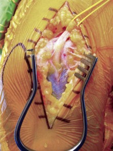
Simple decompression is adequate for the vast majority of patients and can be performed through a relatively small incision that preserves the vascular bed of the nerve. The ulnar nerve is identified proximal to the elbow on the medial aspect of the arm. It can be felt as a rubbery cord. Having mobilized the skin edges, the nerve can be released for 10–15 cm proximal to the skin incision using appropriately angled retractors. Osborne’s fascia is then released between the two heads of the FCU (Fig. 31.5). In simple decompressions the medial intermuscular septum is not excised or released. Having completed the decompression the tourniquet is released, bleeding controlled and the wound closed with a subcuticular suture.

Figure 31.5 (A) Intermuscular septum. (B) Intermuscular septum held. (C) Intermuscular septum incised. (D) Intermuscular septum excised.
In our experience, many of the ulnar nerves that we have successfully decompressed, with complete resolution of symptoms, have a relatively normal appearance at the time of surgery. The normal intraneural vascular markings are easily identified along the length of the nerve and if the tourniquet is released prior to closure, the nerve returns to its deep pink colouration. However if the compressed nerve has become narrowed, fibrosis in the epineurium and between the individual fascicles reduces the chance of a successful surgical outcome.
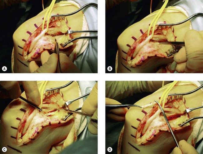
Subcutaneous anterior transposition
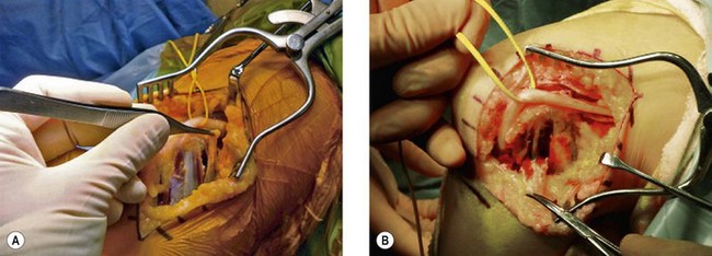
Having positioned the patient and undertaken the incision as previously described the ulnar nerve is identified as it passes beside the medial intermuscular septum. A 4 cm strip of the septum is then excised in order to allow the nerve to remain in the anterior compartment once it has been transposed. Distally the ulnar nerve is released for 5 cm as it passes down between the two heads of the FCU. The nerve can then be mobilized from its bed, preserving its longitudinal blood supply. The new bed for the ulnar nerve is then prepared. The fat is mobilized off the common flexor fascia for approximately 8–10 cm anterior to the medial epicondyle. A fascial strip is then elevated, as shown in Figure 31.6A. The nerve is transposed anteriorly and the fascial strip (Fig. 31.7) looped around the nerve and sutured back onto itself, securing the nerve in its anteriorly transposed position (Fig. 31.8). It is of vital importance that the ulnar nerve does not travel through any sharp angles, proximally, distally or underneath the fascial loop. In particular, the fascial loop should be loose and not cause any compression of the nerve. The transposed nerve should be tension free. The tourniquet is released, haemostasis achieved and the wound closed with a subcuticular suture.

Figure 31.6 (A) Fascial strip fashioned from flexor pronator expansion. (B) Fascial strip dissected to fashion sling.

Stay updated, free articles. Join our Telegram channel

Full access? Get Clinical Tree

