Clinical: Posttraumatic type: painful enlarging mass with intense inflammation of the surrounding tissues. If the involved anatomic compartment is immobilized, inflammation resolves, and the mass slowly regresses. Complete maturation of the mass occurs in 6–12 months. Genetic type: progressive and diffused ossification of tendons, ligaments, and connective tissue around muscles, micro-clinodactyly.
Localization: The posttraumatic type may develop in any site, but it is more frequent in the anterior thigh, upper arm (brachialis muscle), and adductor or gluteal muscles. The genetic type usually starts in the neck, shoulder, axilla, or paraspinal muscles.
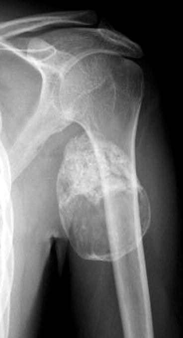
Imaging: Radiological appearance is related to the phase of the process: in the early period only a fading round radiodensity is shown; a peripheral rim becomes visible in few weeks, then the osseous radiodensity enlarges its borders, but a radiolucent central zone remains for a long period. Remodeling of the area occurs for years.
Histopathology: Early phase: spindle cell proliferation, high mitotic activity with hyperchromatic nuclei (d. d. with sarcomas). Then, the peripheral mature and the central immature zone are easily distinguished (zonation phenomenon): loosely arranged spindle cells in center with bone formation at periphery. Mitotically active but without true cellular atypia.
Course: Posttraumatic type: self-limited after rest and anti-inflammatory drugs. Genetic type: progressive severe disability. If respiratory muscles are involved, disease may be fatal.
Treatment: No surgical treatment is usually required. In genetic diseases, good general care and avoidance of trauma (particularly the iatrogenic ones from im injections, biopsies, surgery) are emphasized.