1
Musculoskeletal structure and function

The locomotor system is composed of bone, cartilage, muscle, tendons and ligaments.
Bone
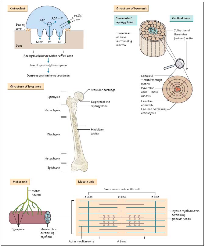
Bone is essentially a mineralised connective tissue. It is comprised of two subtypes:
- Cortical or compact bone comprises 80% of the skeleton, accounting for most of the shafts of long bones. It is formed by Haversian systems: rings of collagen and matrix containing central blood vessels and lining cells called osteocytes.
- Trabecular or medullary bone is found in contact with bone marrow cells between the cortices, at the end of long bones and in vertebral bodies. In trabecular bone the collagen and matrix run as sheets (lamellae) parallel to the bone surface.
The three main cell types in bone are:
Osteoblasts and osteoclasts are coupled into bone remodelling units that keep adult bone mass relatively constant.
See Chapter 31 ‘Disorders of Bone Metabolism’ for more details of osteoclast/osteoblast cell biology.
Bone is covered in a vascular membrane (the periosteum) which is a major source of blood supply to the bone (the other supply is derived from perforating vessels which then run up in the medulla). The periosteum is helpful when reducing fractures, as it is often partly intact and can be used to guide the broken frgments together. The periosteum is also important in fracture healing, supplying cells which ‘organise the haematoma’ around the fracture site (see below).
The protein matrix of bone consists largely of type I collagen. Osteoblasts lay down triple helices of type I collagen into organised lamellae containing unmineralised matrix (osteoid). The tensile strength of bone is increased by covalent bonds between collagen sheets; rigidity is conferred by mineralisation of bone matrix, with deposition of hydroxyapatite crystals between the lamellae.
Bone remodelling occurs throughout life to repair damaged bone. Alternating cycles of recruitment, differentiation and activation of osteoclasts and osteoblasts maintain the structural integrity of bone throughout life; with advancing age however, bone loss exceeds formation. Vigorous bone remodelling follows fracture in 5 stages:
Stay updated, free articles. Join our Telegram channel

Full access? Get Clinical Tree


