10
Anatomy of the spine
Function
The spine is the axial skeleton. It supports the head and the upper limbs, acts as a posterior strut for the thorax and abdomen and at its base locks into the pelvic girdle. It also acts as the conduit for nerve fibres travelling to and from the brain to the rest of the body.
Upper and lower motor neuron lesions
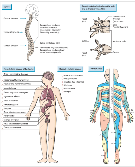
The upper part of the spine (above the first lumbar vertebra) also contains the spinal cord (not just roots) and so is an extension of the central nervous system. Problems in this part of the spine may produce upper motor neuron signs with up-going plantar reflexes and spasticity. Below the first lumbar vertebra, obstruction of the spinal canal can only produce lower motor neuron signs, weakness and wasting combined with sensory loss.
Curves
The healthy spine has three natural curves:
These curves are in part a result of the shape of the bones making up the spine, but are also maintained by the tone of the muscles around the spine. Loss of, or indeed exaggeration of, these curves is suggestive of an underlying disease process.
Vertebrae
Related posts:

Stay updated, free articles. Join our Telegram channel

Full access? Get Clinical Tree


