Microfracture for Chondral Lesions
Michael A. Terry MD
J. Richard Steadman MD
William G. Rodkey DVM
Karen K. Briggs MBA, MPH
History of the Technique
The microfracture technique was initially developed by the senior author (JRS) in the mid-1980s to treat full thickness cartilage defects in the knee. These defects are common and have been shown rarely to heal spontaneously.1,2 The microfracture technique has been modified to its current format over the past 20 years and has been used to treat cartilage lesions in the hip, talus, elbow, and shoulder.3,4,5,7 The procedure began with puncture holes created in exposed bone with a simple awl. The most dramatic modification in technique came after animal studies revealed the importance of removal of the calcified cartilage layer.8 The other significant modifications also involve bed preparation, as discussed below. The rehabilitation protocol has remained essentially the same aside from minor modifications and is also described below.
Indications and Contraindications
Indications for microfracture chondroplasty include full thickness articular cartilage defects in the weight-bearing area between the tibia and femur or in the contact area between the patella and femoral condyles.9,10 Unstable cartilage flaps in these regions that extend to the subchondral bone are also lesions that are suitable for the microfracture technique. Microfracture can be performed on any size lesion, but Steadman et al.10 reported trends of better results with lesions smaller than 400 mm2 without statistical significance. It is preferable, but not required, to have articular cartilage surrounding the lesion that forms an appropriate border for the lesion rather than a lesion that gradually transitions to a full thickness defect. These borders provide some degree of protection and containment for the marrow elements and clot that provide the repair tissue.
Patients with acute injuries to the knee that result in full thickness cartilage loss are treated as soon as appropriate and practical. Patients with chronic lesions or degenerative lesions in the knee are treated conservatively initially for a period of at least 12 weeks. Conservative management for these types of lesions includes: activity modification, physical therapy, nonsteroidal anti-inflammatory medications, and joint injections, as appropriate. Patients who have failed conservative treatment for chronic or degenerative lesions then become candidates for microfracture.
Microfracture should only be performed in patients who are able and willing to undergo the specified postoperative rehabilitation described below. Patients who are unreliable or are unable to complete the rehabilitation required are less likely to benefit from the procedure. Another relative contraindication is malalignment, which could leave the regenerating region under inappropriately high stress during the early phase of repair when the tissue is less durable.11 Advanced age can be a relative contraindication if completion of the rehabilitation protocol is not possible, and it has been demonstrated that age is significant in outcome after microfracture.9,12 Generalized degenerative changes, inflammatory arthritis, and unstable knees are also relative contraindications. None of these are absolute contraindications because of the low morbidity associated with the procedure, but appropriate patient expectations must be created before the procedure is undertaken.9,10,12,13,14
Surgical Techniques
Microfracture can be performed on patients with either regional or general anesthesia. Microfracture under local
anesthesia with sedation has not been studied and should be used with care because of the potentially adverse environment created by the local analgesia.
anesthesia with sedation has not been studied and should be used with care because of the potentially adverse environment created by the local analgesia.
We place our patients in a supine position on a standard operating room table for knee arthroscopy. We apply a tourniquet but generally do not inflate it for microfracture procedures.
Standard arthroscopic portals can be used for microfracture. We make three portals: a superior and medially placed inflow portal and medial and lateral parapatellar portals. Accessory portals are occasionally made as needed for lesions in difficult locations. We perform a standard and thorough diagnostic arthroscopy of the knee prior to microfracture. If other pathology is present in the knee that requires treatment, we complete such treatment prior to microfracture, with the exception of ligament reconstruction. This technique decreases the amount of time that the microfractured bone is exposed to the elevated intra-articular pressures and fluid flow that can decrease the formation of the clot, which is critical to success.
Once a lesion is identified for microfracture (Fig. 58-1), it is probed thoroughly to ensure that all bordering cartilage is stable (Fig. 58-2). It is then debrided of all loose flaps of cartilage using a shaver (Fig. 58-3). Creation of stable full-thickness borders of cartilage surrounding a central lesion is optimal for microfracture as it provides some degree of protection to the regenerating tissue that is forming in the treated lesion. All loose cartilage and cartilage flaps should be removed regardless of border conditions.
The removal of the calcified cartilage layer is important and is usually performed carefully using a hand held curette (Fig. 58-4).8 Care must be taken at this stage. Subchondral bone destabilization or loss of the articular morphology results from overly aggressive debridement. Debridement and bed preparation that are inadequate will leave cartilage on the underlying bone, potentially inhibiting the regeneration process.8
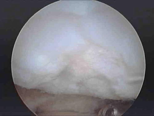 Fig. 58-1. Cartilage defect is identified and selected for microfracture. |
 Fig. 58-2. The edges of each defect selected for microfracture must be thoroughly probed to ensure that the cartilage along the border is stable. |
 Fig. 58-3. Loose flaps of cartilage are removed using a motorized shaver.
Related posts: Open Anterior Instability Repair
Rotator Cuff Repair Part I. Arthroscopic Approach
Acromioclavicular Reconstruction Using a Free Tendon Graft and Interference Screw Fixation
Miscellaneous
Medial and Lateral Epicondylitis: Open Treatment
Management of the Arthritic and Stiff Elbow and Osteochondritis Dessicans of the Capitellum Open Anterior Instability Repair
Rotator Cuff Repair Part I. Arthroscopic Approach
Acromioclavicular Reconstruction Using a Free Tendon Graft and Interference Screw Fixation
Miscellaneous
Medial and Lateral Epicondylitis: Open Treatment
Management of the Arthritic and Stiff Elbow and Osteochondritis Dessicans of the Capitellum
Stay updated, free articles. Join our Telegram channel
Full access? Get Clinical Tree
 Get Clinical Tree app for offline access

|