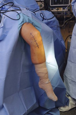
Chapter 85 Injury to the medial aspect of the knee has traditionally been thought of as simply a medial collateral ligament (MCL) injury, but more recent research has concluded that the main medial knee structures have a complex relationship and provide both static and dynamic stability. The majority of acute isolated medial knee injuries can be treated nonoperatively, and a full return of function should be expected. The treatment of injuries to the medial knee had undergone a shift from aggressive operative treatment to almost exclusively nonoperative treatment; however, with the advent of new reconstructive techniques and acute augmentation techniques, the pendulum has swung back toward operative treatment in select patients.1 The surgical reconstruction of choice should recreate the native anatomy and be justified by both biomechanical and clinical research. The patient with a medial-sided knee injury can usually recall the injury mechanism. This typically is a valgus load to the knee and can occur when the foot is planted or from a direct lateral blow.2 The recollection of hearing a “pop” can hint at the presence of a cruciate ligament injury or bone bruise. In the chronic setting, patients will describe the feeling of a side-to-side instability. Localized swelling and induration are present around the medial femoral condyle and/or proximal tibia. Deep medial joint line tenderness specifically is usually found only in the setting of a concomitant meniscal injury. The presence of a large hemarthrosis should alert the examiner to a potential combined cruciate ligament injury. The medial structures should be tested by the application of a valgus load at both full extension and 20 degrees of flexion. Isolated medial knee injuries will gap in 20 degrees of flexion, but the presence of gapping at full extension should heighten the suspicion for a concurrent cruciate ligament injury.3 Of note, in our experience the presence of gapping in full extension at time zero (acute injury) is associated with an increased risk of failure of nonoperative treatment and chronic valgus laxity despite cruciate ligament reconstruction. The medial knee injury can be classified into three grades. Grade I injuries indicate a partial tear of the MCL and, on valgus stress, subjectively gap less than 5 mm. A grade II injury subjectively gaps 5 to 9 mm with valgus stress at 20 degrees of flexion and does not gap in extension.4 Both of these injuries have firm endpoints with a valgus load. A grade III injury indicates a complete tear of the MCL. This injury subjectively gaps more than 10 mm at 20 degrees of flexion and often gaps at full extension as well. The examiner will typically find a soft or no endpoint. A thorough ligamentous examination should be performed, including an anterior drawer, posterior drawer, Lachman, pivot-shift, reverse pivot-shift, dial, and posteromedial, anteromedial, and posterolateral drawer tests. It is very important to note that a positive dial test does not always indicate a posterolateral corner injury. Complete injury to the medial structures significantly increases external rotation at 30 and 90 degrees of flexion, resulting in a positive dial test.5 It often occurs that a chronic medial knee injury will be misdiagnosed as a posterolateral corner injury owing to the presence of a positive dial test. The injured and uninjured knees should be imaged with standard anteroposterior, lateral, patellofemoral, Rosenberg, and standing long-leg radiographs. Specifically, films should be evaluated for fractures, loose bodies, and capsular or ligamentous avulsions. In the chronic setting, valgus stress radiographs should be obtained to quantify the amount of medial compartment opening and should be compared with radiographs of the uninjured knee. If there is confusion regarding the exact diagnosis (positive dial test result), varus stress radiographs can be obtained as well. Gapping of more than 3.2 mm at 20 degrees of flexion is indicative of a grade III superficial MCL injury, whereas gapping of 6.5 mm at 0 degrees and 9.8 mm at 20 degrees of flexion is consistent with a complete medial-sided knee injury with rupture of the superficial fibers of the medial collateral ligament (sMCL), deep MCL, and posterior oblique ligament (POL).6 Magnetic resonance imaging (MRI) should be performed to evaluate for meniscal, cartilaginous, or other ligamentous injury and the presence of a distal meniscotibial Stener lesion, which might indicate the need for operative intervention.7 The presence of lateral femoral condyle and lateral tibial plateau bone bruises should alert the physician to the possibility of a medial-sided injury. These lesions have been reported in 45% of medial knee injuries and typically resolve within 4 months from the time of injury.8 Three main structures provide stability to the medial knee—the sMCL, the deep MCL, and the POL. These structures provide restraint to valgus, external rotation, and internal rotation forces. The sMCL is the primary restraint to valgus load on the knee. It averages 9.5 cm in length and has one femoral and two tibial attachments. The femoral attachment site is not to the medial epicondyle, but is 3.2 mm proximal and 4.8 mm posterior to the medial epicondyle.9 The proximal tibial attachment is 1.2 cm distal to the joint line and is primarily to soft tissues (anterior arm of the semimembranosus). The distal tibial attachment is reproducibly 6 cm distal to the joint line regardless of the patient’s size. The proximal division of the sMCL is important for valgus stability, whereas the distal division is more important in stabilizing external rotation. The patient is placed supine on the operating table, and general anesthesia is induced. A preoperative femoral nerve indwelling catheter is placed by the anesthesiologist under ultrasound guidance, and 0.25% bupivacaine is administered for approximately 48 hours postoperatively to assist in postoperative pain control. The nonoperative leg is placed into a well-leg holder, and the fibular head is well padded. A thorough examination under anesthesia is performed, including evaluation of range of motion and the ligamentous examination described earlier. A nonsterile tourniquet is applied to the operative extremity, and the limb is placed into an arthroscopic leg holder after the foot of the bed has been completely lowered (Fig. 85-1). A surgical time-out to verify the operative extremity is performed, and prophylactic antibiotics are administered. The limb is prepared with chlorhexidine and draped in the standard fashion.
Medial Collateral Ligament and Posteromedial Corner Repair and Reconstruction
Preoperative Considerations
Physical Examination
Imaging
Magnetic Resonance Imaging
Anatomy
Surgical Technique
![]()
Stay updated, free articles. Join our Telegram channel

Full access? Get Clinical Tree


Medial Collateral Ligament and Posteromedial Corner Repair and Reconstruction


