Measles (Rubeola)
Andreea C. Cazacu
Gail J. Demmler
Infection with measles virus produces an illness characterized by a prodrome of fever, coryza, cough, conjunctivitis, an enanthem (Koplik spots), and development of a confluent, erythematous maculopapular rash. The mortality rate associated with measles is approximately 1 in 3,000 cases in the United States. A higher mortality and increased morbidity is seen in young infants, immunocompromised children, and pregnant women. In developing countries, such as Africa, mortality rates of 10% or higher occur in malnourished children who contract measles.
Measles epidemics were described in both the Roman Empire and ancient China. By the seventeenth century, differentiation between measles and smallpox was made, and reports of measles in London and colonial America were described. In the late 1800s, the pathognomonic enanthem of measles was reported in detail by Koplik.
In 1954, Enders and Peebles isolated measles virus in human and simian tissue culture lines. Cultivation of measles virus in chicken embryo tissue enabled vaccine development to proceed. Vaccine trials occurred in the late 1950s and early 1960s, with vaccine licensure in 1963.
ETIOLOGY
Measles virus, which has an internal core of RNA and an outer envelope of glycoproteins and lipids, is a member of the family of Paramyxoviridae, genus Morbillivirus. Two glycoproteins, hemagglutinin (H) and fusion (F), are important in immune protection responses. Immune responses to these two glycoproteins include hemagglutination antibody response in infected
individuals (H) and fusion of nucleated cells with formation of multinucleated giant cells (F). Measles virus is monotypic, sensitive to both heat and cold, and inactivated by ultraviolet light.
individuals (H) and fusion of nucleated cells with formation of multinucleated giant cells (F). Measles virus is monotypic, sensitive to both heat and cold, and inactivated by ultraviolet light.
EPIDEMIOLOGY
Measles is a highly contagious disease and occurs throughout the world as a winter–spring disease in temperate climates. Person-to-person transmission of the measles virus occurs by droplet spread of respiratory secretions with acquisition of infection by the nasopharyngeal route and possibly the conjunctivae. The highest period of infectivity is the catarrhal period before appearance of the exanthem. Person-to-person spread of measles occurs in the home among family members, in medical settings between patients and health care workers, and among students and teachers in day-care centers, schools, colleges, and universities.
In the prevaccine era, highest attack rates were seen in children 5 to 10 years of age, although in urban populations, a higher incidence was observed in preschool-aged children. In developing countries, the highest attack rates occur in children 2 years of age or younger. When the widespread use of measles vaccine began in 1965, the incidence of measles fell remarkably and in 1983 reached an all-time low. In 1986, however, the incidence of measles increased and in 1989 and 1990 reached epidemic proportions in many areas of North America. This increase in incidence of measles was most striking among unvaccinated preschool-aged children, adolescents, and young adults, especially those individuals who were foreign-born. However, vaccine failure, occurring in as many as 5% of individuals who received one dose of measles vaccine, also may have contributed to the observed increase in incidence of measles, and led to current recommendations that children receive two doses of measles vaccine prior to adolescence.
PATHOGENESIS AND PATHOLOGY
Measles virus infection of the nasopharynx respiratory epithelium spreads to regional lymphatics, resulting in viremia. This viremia is enhanced by replication of the virus in the reticuloendothelial system. The respiratory tract, skin, and conjunctivae are major sites of infection, but other organs may be involved. Viral replication peaks in all organs and the blood at approximately the same time that the rash appears, followed by development of immune responses and subsequent curtailment of the illness.
Formation of multinucleate giant cells—the result of cell fusion—characterizes the pathologic response to measles virus infection. These cells are found in the skin, respiratory tract, reticuloendothelial system, and other organs. They contain both intracytoplasmic and intranuclear eosinophilic inclusions.
Immune responses to infection with measles include hemagglutination inhibition (HAI) and production of neutralizing and complement fixing (CF) antibody. In natural infection, antibody responses appear at approximately 14 days and peak several weeks later, with a range of 4 to 6 weeks. The CF antibody appears later than HAI and does not usually persist. The IgM antibody response appears early in the illness, but rarely persists beyond 90 days.
Infected hosts develop cell-mediated immunity and interferon response in the serum. Delayed hypersensitivity responses are suppressed by infection with natural measles virus as well as by vaccine strains. Immunocompromised hosts, especially individuals with T-cell lymphocyte dysfunction, may have a prolonged course with measles, a prolonged duration of excretion of virus, and a high incidence of morbidity and mortality.
CLINICAL MANIFESTATIONS
Persons with measles virus infections fall into four distinct clinical groups: typical measles in the normal host; modified measles in a host with preexisting antibody; atypical measles in the host who received killed vaccine; and measles in immunocompromised, malnourished, or special hosts.
Typical Measles
The incubation period in typical measles is approximately 10 days, starting with a 3- to 5-day prodrome of malaise, fever, cough, coryza, and conjunctivitis. These symptoms increase over the 3- to 5-day prodrome period. Fever ranges from 39.4°C to 40.6°C, reaching its highest at the nadir of exanthem. About 2 days before the rash, Koplik spots (white, pinpoint lesions on a bright red buccal mucosa) appear first opposite the lower molars and quickly spread to involve the entire buccal and lower labial mucosa. Koplik spots resolve by the third day of exanthem. The exanthem of measles starts about the fourteenth day after exposure and appears first behind the ears and the hairline of the forehead. The rash progresses downward to the face, neck, upper extremities, and trunk and reaches the lower extremities by the third day. Initially, the rash is discrete, erythematous, and maculopapular, but it becomes confluent in the same progression as its spread (Fig. 202.1). Eventually the rash undergoes a brownish discoloration that does not blanch with pressure and may desquamate. The exanthem lasts 6 to 7 days, and resolution of the rash proceeds in the same order as that of its appearance. In uncomplicated measles, fever resolves in the first week; increased temperature beyond the third or fourth day of the exanthem suggests a complication.
Pharyngitis as well as generalized adenopathy may be seen during the period of exanthem. Splenomegaly is a common occurance. Diarrhea, vomiting, and abdominal pain may be prominent symptoms of measles, especially in young children. Leukopenia is a predictable finding.
Modified Measles
Modified measles occurs in children who have received immune serum globulin or intravenous immune globulin
preparations, or in very young infants who still have transplacentally acquired maternal measles antibody. In addition, vaccine-modified mild measles, a form of secondary vaccine failure, can occur in individuals who were appropriately vaccinated with the live measles virus vaccine. In mild or modified measles, the prodrome period is shortened, symptoms are not as severe, and Koplik spots usually do not occur, and if present, they fade rapidly. The exanthem follows the progression of regular measles but appears faint and does not become confluent.
preparations, or in very young infants who still have transplacentally acquired maternal measles antibody. In addition, vaccine-modified mild measles, a form of secondary vaccine failure, can occur in individuals who were appropriately vaccinated with the live measles virus vaccine. In mild or modified measles, the prodrome period is shortened, symptoms are not as severe, and Koplik spots usually do not occur, and if present, they fade rapidly. The exanthem follows the progression of regular measles but appears faint and does not become confluent.
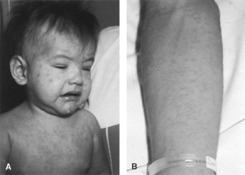 FIGURE 202.1. Maculopapular rash. A: Typical measles. (Courtesy of Dr. Gail J. Demmler.) B: Atypical measles. See Color Figure 202.1 in color section. |
Atypical Measles
Atypical measles is rarely seen today. It occurred in persons immunized with killed measles virus vaccine who were exposed to natural measles. This illness may be observed today in older adults who received killed measles vaccine from 1963 to 1967 and were not reimmunized with live virus vaccine.
The incubation period for atypical measles is the same as for typical measles. The illness is characterized by sudden onset of fever (39.4°C to 40.6°C). Headache, myalgias, extreme weakness, and abdominal pain all may be present. Almost all patients have a dry, nonproductive cough.
The rash of atypical measles appears first on the distal extremities and is pronounced on the wrists and ankles (see Fig. 202.1). The rash may remain localized or spread to involve the upper and lower extremities as well as the trunk. The palms and soles are also involved. The rash of atypical measles typically is erythematous and maculopapular; however, it also may be vesicular, petechial, or purpuric in nature, and urticaria, edema of the hands and feet, and severe hyperesthesia also have been described. Koplik spots are rarely seen.
Related posts:

Stay updated, free articles. Join our Telegram channel

Full access? Get Clinical Tree


