Managing Complications Following Hip Resurfacing
Thomas Parker Vail
Abstract
Metal-on-metal hip resurfacing arthroplasty (HRA) has been very successful in selected patients at experienced centers. However, revision rates of modern metal-on-metal HRA implants have been reported at rates from as low as 2% at 5 years up to 20%. The most commonly reported mechanisms of failure of modern hip resurfacing include femoral neck fracture, femoral component loosening, and failure of acetabular component fixation. Less commonly reported failure mechanisms of hip resurfacing include infection, extra-articular impingement or other soft tissue–related causes of persistent groin pain, and complications of metal ion exposure or unintended wear. Retrieval analysis of failed implants reveals mechanical factors such as edge loading associated with certain failure modes. Histologic findings from tissues retrieved at revision surgery can show a variety of findings related to the mechanism of failure including osteonecrosis, soft tissue inflammation and necrosis, and lymphocytic infiltration of soft tissue. Careful evaluation for adverse mechanical or biologic processes will inform the decision to revise or monitor. The most common management of a failed resurfacing implant is conversion to a total hip replacement.
Case Presentation
The subject is a 59-year-old male with a history of osteoarthritis of the left hip. His symptoms were resistant to medical management. After a review of treatment options, he opted to undergo a metal-on-metal total hip resurfacing procedure. Surgery was performed by an experienced surgeon without complications in 2004. The patient recovered uneventfully from surgery and resumed an active lifestyle including walking, swimming, and biking.
In 2012 he presented in the clinic complaining of nagging groin pain with activities of daily living that had been present for several years. He reported that the surgery never completely eliminated the pain in his hip although he had periods of time with very little pain. Over the past 6 months prior to presentation the pain and limp had been getting progressively worse, causing him to use a cane intermittently and limiting walking to less than 30 minutes. He was no longer able to tolerate swimming or stationary bicycle. Physical examination revealed a slender, healthy appearing male with a mildly antalgic gait, no Trendelenburg sign, and no palpable crepitus or soft tissue fullness. An antero-posterior radiograph of his hip (Fig. 69.1) shows a total hip resurfacing implant with a slightly vertical socket and a noncontinuous retroacetabular radiolucency noted on the radiographs. Laboratory studies included an ESR of 1 mm/hr (nl range 0 to 10 mm/hr) and CRP of 1.6 mg/L (nl <1 mg/L). An outside bone scan report indicated “mild uptake around the acetabular component.”
Introduction
Hip resurfacing arthroplasty (HRA) is a femoral head bone–conserving alternative to total hip arthroplasty that is indicated in the clinical setting of end-stage hip arthrosis with femoral head bone that is structurally adequate to support a femoral resurfacing implant (1). Resurfacing at this time is largely limited to metal-on-metal constructs, although in the past other polymer and ceramic materials as well as soft tissue interposition have been used for the same purpose (2). The ideal candidate for HRA is being defined and redefined by analysis of clinical outcomes in the published worldwide experience (3,4,5,6,7,8,9,10,11), international joint registries, and retrieval studies (12,13,14,15). While the final decision on indications for hip resurfacing surgery remains in the domain of the individual doctor and patient using shared decision making, the best aggregate results have been reported in younger males of larger stature (16,17). Revisions have been more common in smaller individuals, older patients, and females. In addition, concerns have been raised regarding potential adverse effects on local tissues or systemically from metal ions derived from the metal-on-metal bearing.
While the name “surface replacement” implies a simple concept, the procedure is technically demanding, requiring
skill in implantation of a monoblock cobalt-chrome socket and a metallic resurfacing femoral component. The surgical approach requires knowledge of the blood supply to the femoral head, protection of the soft tissues to avoid compromising the blood supply to the femoral head, and adequate exposure to achieve desired implant position (2,18). There are many examples of outstanding results in the hands of experts published in reports of case series and the larger experience of all hospitals and surgeons in several national joint replacement registries throughout the world. However, the technical challenges and variability of outcome are also apparent from the same body of literature. For example, the initial clinical experience with a larger cohort of patients and multiple surgeons in the United States (19,20,21) does not mirror the success reported by experienced experts in single institution case series.
skill in implantation of a monoblock cobalt-chrome socket and a metallic resurfacing femoral component. The surgical approach requires knowledge of the blood supply to the femoral head, protection of the soft tissues to avoid compromising the blood supply to the femoral head, and adequate exposure to achieve desired implant position (2,18). There are many examples of outstanding results in the hands of experts published in reports of case series and the larger experience of all hospitals and surgeons in several national joint replacement registries throughout the world. However, the technical challenges and variability of outcome are also apparent from the same body of literature. For example, the initial clinical experience with a larger cohort of patients and multiple surgeons in the United States (19,20,21) does not mirror the success reported by experienced experts in single institution case series.
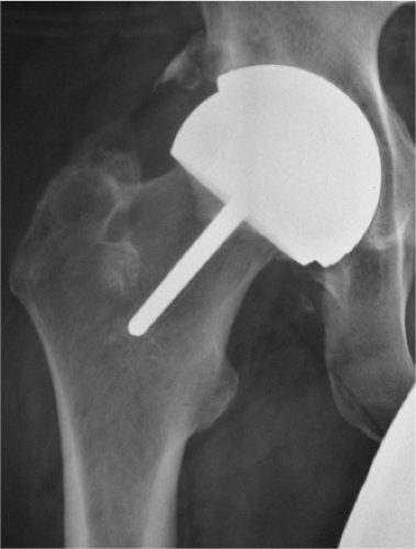 Figure 69.1. HRA with a vertical socket but no obvious loosening in a patient with groin pain at 6 months after surgery. |
Specific technical challenges of HRA include acetabular component seating, primary stability of the implants, acetabular cup position, femoral component seating and positioning, protection of the femoral neck cortex (18), and protection of the femoral head and neck blood supply (22). Thus, the technical challenges in HRA are different from total hip replacement in many regards (1,23,24). Attention to specific details unique to the technique of hip resurfacing can enhance the probability of a successful outcome of the procedure. For example, Amstutz (25,26) and others (27) have found that changes in cement technique can lead to lower rates of thermal necrosis of bone and component loosening.
Complications or early failures can sometimes be related to technical issues and patient selection (18,28,29). Managing complications of hip resurfacing requires knowledge of a unique profile of complications derived from the technical challenges that can lead to a decision to revise. Femoral neck fracture is the most common early complication of hip resurfacing (30,31,32,33) followed by component loosening, infection, unexplained pain, and adverse tissue reactions. In a review of a consecutive series of resurfacing implant retrievals, Morlock et al. (13,14) noted that 20 of the 281 retrievals were complete bearing revisions (both the femoral and acetabular components of the bearing couple). The most frequent reason for revision was fracture of the femoral neck initiated at the rim of the resurfacing implant (42.5%) after an average of 122 days, followed by fracture originating from inside of the resurfacing component (17.6%) after 267 days. However, only 60% of the revisions for fracture were due to acute fractures of the femoral neck. Revision of the femoral component due to loosening without an acute fracture (28%) occurred after nearly 2 years (700 days) followed by acetabular cup loosening (11.9%), which occurred after an average of 460 days.
While it is not realistic to eliminate all early failures, some early complications of hip resurfacing leading to reoperation and revision are potentially avoidable. In particular, the failures associated with technical factors could be minimized by application of experience and knowledge and careful patient selection (34). Without doubt, some complications of hip resurfacing such as osteonecrosis (7,35) or adverse soft tissue reaction to metal particles may not be entirely avoidable even with experience and optimization of technique (36,37,38). Some have suggested that protecting the blood supply to the femoral head may provide some protection against osteonecrosis of the bone after hip resurfacing (39,40). Clinical experience suggests that complete vascular disruption is not an inevitable consequence of femoral head resurfacing (41). Postoperative PET scanning also supports the notion that femoral head viability can be maintained after hip resurfacing (42).
The specific aim of this chapter is to review the routine follow-up of hip resurfacing patients, the approach to the patient with elevated metal levels or unexplained pain, the management of the failed femoral component due to loosening or fracture, the management of the failed acetabular component secondary to failed ingrowth or malposition, and finally the surgical management of the patient who has associated soft tissue damage.
Routine Follow-Up and Evaluation of the Painful Hip Resurfacing Implant
Routine evaluation (43) of a hip resurfacing is similar to the routine evaluation and follow-up of any arthroplasty patient with particular diligence to detect problems related to fixation, infection, or bearing function. For most patients, the return to full function can be expected within 3 months of the procedure, acknowledging that this period of time may vary depending upon the patient and any unique circumstances. Likewise, it is not uncommon to see subtle improvements in motor function, strength, coordination, and athletic ability
extend up to 1 year after surgery. Using that general description as a metric, any patient who falls off of that expected course of recovery should be evaluated. Radiographs of the proximal femur and pelvis are important first-line components of regular evaluation and follow-up in conjunction with the clinical history and physical examination. In scheduling follow-up, the care providers should keep in mind the following time sequence of potential complications: wound healing or acute infection occurs within the first weeks of surgery, femoral neck fracture is most common within the first 3 months, early implant loosening or failure of acetabular bone ingrowth may be manifest by a patient who complains of groin pain from the outset of recovery and never improves, femoral component loosening generally occurs after the first 6 months, chronic soft tissue pain (iliopsoas impingement or capsular impingement) not related to pain associated with the initial healing process may be manifest after 6 months, and complications related to metal ions are generally not apparent until after 6 months but can manifest years after successful surgery (Table 69.1). Thus, routine follow-up should be designed to assess proper wound healing and rehabilitation in the initial stages, evaluate lingering pain after the expected period of acute recovery, and detect early manifestations of bearing problems known to arise later in the postoperative course.
extend up to 1 year after surgery. Using that general description as a metric, any patient who falls off of that expected course of recovery should be evaluated. Radiographs of the proximal femur and pelvis are important first-line components of regular evaluation and follow-up in conjunction with the clinical history and physical examination. In scheduling follow-up, the care providers should keep in mind the following time sequence of potential complications: wound healing or acute infection occurs within the first weeks of surgery, femoral neck fracture is most common within the first 3 months, early implant loosening or failure of acetabular bone ingrowth may be manifest by a patient who complains of groin pain from the outset of recovery and never improves, femoral component loosening generally occurs after the first 6 months, chronic soft tissue pain (iliopsoas impingement or capsular impingement) not related to pain associated with the initial healing process may be manifest after 6 months, and complications related to metal ions are generally not apparent until after 6 months but can manifest years after successful surgery (Table 69.1). Thus, routine follow-up should be designed to assess proper wound healing and rehabilitation in the initial stages, evaluate lingering pain after the expected period of acute recovery, and detect early manifestations of bearing problems known to arise later in the postoperative course.
Table 69.1
Get Clinical Tree app for offline access
Related posts:Stay updated, free articles. Join our Telegram channel
Full access? Get Clinical Tree


|
|---|


