Leg, Ankle, and Foot: History, Physical, and Investigations
Kevin J. Wing MD, FRCSC
Alastair Younger MSC, MBCHB, FRCSC
By the end of the history, the examiner should know the functional restrictions of the patient and the specific areas of discomfort. The nature of the injury should be determined, if an isolated event. Insidious onset may be related to change in routine.
Changes in training routine or footwear can aggravate or cause a stress injury.
In an elite female athlete, it is appropriate to inquire about menstrual history, keeping in mind the female athlete triad of disordered eating, amenorrhea, and osteoporosis.
Determination of the absolute range of motion is less critical than assessment of loss of motion compared with the opposite side, as motion is very variable between patients.
The recent development of Multidetector-row computed tomography (CT) has dramatically improved the quality of foot and ankle images. The high resolution axial scans, in conjunction with sagital and coronal reformats, has improved the diagnosis and visualization of osteochondral lesions of the talar dome, arthritic joints, stress fractures, or small rim fractures.
We have added this chapter on the examination of the foot and ankle in the athlete. Most errors in the management of foot and ankle conditions in the athlete are caused by a poor history and physical, and the examination is not as well taught or thought out compared with other areas of the body. We hope you will learn from inclusion of this chapter.
By the end of the foot and ankle history, the examiner should know the functional restrictions of the patient and the specific areas of discomfort. The nature of injury should be determined, if an isolated event. Insidious onset may be related to changes in routine. Patients typically report one or a combination of pain, instability, and locking.
Pain
Patients should remove their socks and shoes prior to the history so they can indicate the area of discomfort or deformity. This seems to be a much more effective method of communication than verbal description.
Patients should describe the severity of their pain, as well as the quality, radiation, and timing. They should also describe any precipitating and relieving factors. The effect of hills and stairs should be determined (anterior and posterior ankle impingement). Subtalar or triple joint pathology and peroneal tendon problems may cause pain on uneven ground. The effects of anti-inflammatories or acetaminophen on their pain pattern should also be noted.
Instability
Instability most commonly affects the ankle joint. It is the result of laxity of the anterior talofibular ligament (ATFL). The calcaneal fibular ligament (CFL) is also frequently found to be deficient at the time of surgery.
The examiner should delineate between apprehension and true inversion sprains. The frequency of these episodes and the setting in which they occur should be assessed.
Locking
Locking in the foot and ankle may relate to an osteochondral defect or loose body within the ankle. Patients may find the symptoms hard to define compared to pain and instability.
Functional Restriction
The impact of the disability should be determined. Sports practiced prior to and since the onset of symptoms are recorded. Impact on specific sports and activities within sports (running, cutting, jumping, and so on) will determine the need for treatment.
Impact on employment should also be recorded.
Treatment to Date
Treatment to date may include physiotherapy, medication, surgery, bracing, or orthotics. Knowledge of prior treatment and its success or failure will allow the physician to plan an appropriate treatment strategy. For instability, specific treatment may include bracing and physiotherapy. Patients should bring in their braces and orthotics for review.
Change in Foot Shape
The onset of an acquired flat foot deformity or an acquired post-traumatic flat foot deformity is an important finding in the adult. We have found a series of patients with superficial deltoid injuries presenting with a planovalgus foot deformity. Patients with midfoot injuries (Lisfranc joint level) may also develop a flat foot deformity.
A progressive high arched (cavovarus) foot deformity is less common but significant symptom often associated with instability. This pertains more to the younger athlete encountering a neuromuscular disorder, such as Charcot Marie Tooth. Occasionally patients will develop a high arched foot after an acute injury to the talar neck. This can be missed or the nature of the injury underestimated. A painful locked foot with hindfoot varus and forefoot varus will result.
Previous Foot and Ankle Conditions
Prior injuries, such as high school injuries, may be only volunteered on specific questioning. Pre-existing deformities or family history of flat or high arched feet are also significant. Many of these deformities become more symptomatic with age.
Stress/Overuse Injuries
Changes in training routine or footwear can aggravate or cause a stress injury (1,2). A history of the exercise routine should be sought.
In an elite female athlete it is appropriate to inquire about menstrual history, keeping in mind the female athlete triad of disordered eating, amenorrhea, and osteoporosis (3).
Past Medical History
Recording the typical past medical history, past surgical history, current medications, and allergies must complete the history section. A patient filled form may be the most reliable screening method for obtaining details.
The feet should already be exposed for the history. The height and weight should be recorded. Terminology should accurate: Because pronation and supination mean different things to different practitioners, we prefer to describe feet in general as flat, high arched, or neutral. More specific description of foot shape includes hindfoot varus and valgus, forefoot varus and valgus (inverted or everted toward or away from the midline), and forefoot abduction and adduction in the transverse plane. This is consistent terminology that most practitioners understand.
We use a nine-point physical exam that ensures that all of the relevant details in the physical exam are covered. These points are discussed in the following sections.
Exposure
The physical exam should be conducted with the patient dressed in shorts and a T-shirt, so that the alignment of the limb can be clearly seen (Fig 45-1).
Gait
A brief examination of gait requires a moderate sized exam room or corridor of at least 10 to 15 feet (3 to 5 m). The patient is asked to walk back and forth on three occasions, watching first the left foot and its posture in stance phase. On the return the position, the swing phase is watched. The routine is then repeated for the right foot. The patient is asked to heel walk out and toe walk back.
Inspection of Standing Posture
Standing posture starts with an observation of alignment with the patient facing the examiner and feet shoulder width apart. A “peek-a-boo” heel may be observed indicating hindfoot varus (Fig 45-2) (4). The weight bearing position of the toes (clawing, hallux valgus) is inspected.
After the patient turns around the posture is observed from behind. A quick screening exam of the back, pelvic alignment, and measurement of significant leg length discrepancy can be performed now. Focusing on the foot, the examiner can determine whether the heel is in neutral
(three to six degrees) physiologic valgus. A flat foot will typically be associated with a hindfoot valgus and lateral displacement of the forefoot indicated by the “too many toes” sign (5) (Fig 45-3).
(three to six degrees) physiologic valgus. A flat foot will typically be associated with a hindfoot valgus and lateral displacement of the forefoot indicated by the “too many toes” sign (5) (Fig 45-3).
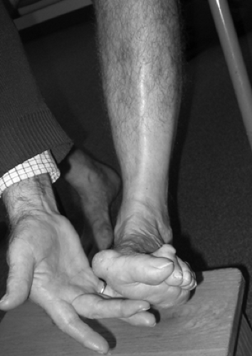 Fig 45-1. Exposure of the limb. The patient is pointing at the painful second metatarsal base. |
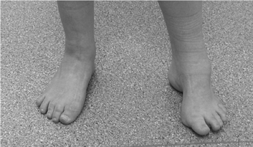 Fig 45-2. Peek a boo heel sign. A patient with Charcot Marie Tooth disease with a reconstructed foot on the right and preoperative foot on the left. The right appears well aligned, and the left has a peek a boo heel. |
A single leg stance will assess proprioceptive control of the lower extremity. This can be made more vigorous by asking the patient to hop while on one leg. The patient can be asked to do a single leg heel raise to both ascertain proprioceptive control in plantarflexion, and that they reconstitute a normal longitudinal medial arch and have normal heel inversion as a result of tibialis posterior tendon function and a supple hindfoot (6).
 Fig 45-3. Too many toes sign. |
Inspection of the Foot with the Patient Sitting
The patient is moved to a sitting position preferably on a slightly raised bed with the legs hanging over the edge and the examiner seated on a low stool. This allows optimal visualization of the leg, foot, and ankle.
The foot is examined for callosities that indicate areas of excessive load in the foot. Common sites include metatarsal heads, the lateral border of the foot (cavus foot), and over the interphalengeal joints (claw toes).
Any scars from previous surgeries or injuries are inspected and recorded. Their effect on any further surgery should be considered. They should be palpated and examined for any signs of infection.
Swelling may exist over a joint or tendon. Its location and extent should be recorded. If necessary, assessment of tibial rotation, length, varus, or valgus alignment may be relevant after fracture or growth arrest.
Palpation
Palpation is the critical part of examination. Determination of the areas of maximum tenderness and knowledge of the underlying anatomy will point to the diagnosis.
Joint swelling and osteophytic or post-traumatic bony changes at the joint margins should be noted.
The following joint margins should be palpated.
Ankle joint: Anterior joint line, medial and lateral gutters (Fig 45-4)
Subtalar joint: Sinus tarsi, middle facet of the subtalar joint
Talonavicular joint
Calcaneocuboid joint
Navicular cuneiform joint
Tarsal metatarsal joints 1–5
Metatarsal phalangeal joints 1–5
IP joints 1–5
DIP joints 2–5
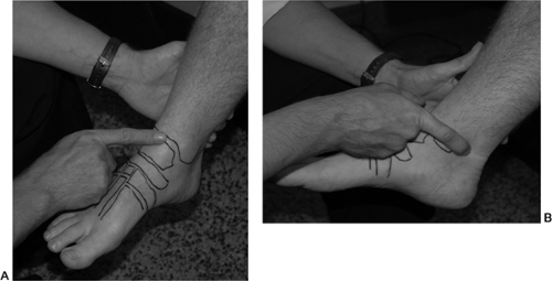 Fig 45-4.A: Anterior ankle joint margin palpation. B: Ankle joint margin palpation—posterior margin.
Related posts: Periarticular Ligamentous Tissue: A Biochemical and Physiologic Assessment
Functional Spinal Rehabilitation
Acute Injuries: Shoulder Fractures and Acromioclavicular and Sternoclavicular Joint Injuries
Hip Arthroscopy
Medial Collateral Ligament
Anatomic Double Bundle Anterior Cruciate Ligament Reconstruction Periarticular Ligamentous Tissue: A Biochemical and Physiologic Assessment
Functional Spinal Rehabilitation
Acute Injuries: Shoulder Fractures and Acromioclavicular and Sternoclavicular Joint Injuries
Hip Arthroscopy
Medial Collateral Ligament
Anatomic Double Bundle Anterior Cruciate Ligament Reconstruction
Stay updated, free articles. Join our Telegram channel
Full access? Get Clinical Tree
 Get Clinical Tree app for offline access

|