Fig. 8.1
Clinical image of intense chronic synovitis in a young hemophiliac patient. The problem was treated by radiosynovectomy with Yttrium-90
In general, it is recommendable to perform a nonsurgical synovectomy before indicating any type of surgical synovectomy, given that the nonsurgical method is much simpler and easier (with similar efficacy). We use radiosynovectomy with Yttrium-90 in children over 12 years of age, and we prefer arthroscopic synovectomy in children under that age. The dose we use for knee radiosynovectomy is 185 MBq. Yttrium-90 is a pure beta-emitter, with therapeutic penetration power of 2.8 mm and an average life of 2.8 days. We have never used chemical synovectomy because it requires multiple painful weekly injections of rifampicin or oxytetracycline.
On the knee, arthroscopic synovectomy requires good perioperative hemostasis, as synovial tissue bleeds a lot. The appropriate clotting control via the infusion (continuous or bolus) of the deficit factor is essential to prevent postoperative bleeding [5]. Arthroscopic synovectomy usually requires at least two entry points in order to resect as much synovial as possible [11]. After synovectomy (of any kind), it is recommended to use a compression bandage for 3–4 days and ensure the joint has limited mobility (that permitted by the bandage).
In summary, radiosynovectomy and arthroscopic synovectomy are effective methods to control recurrent hemarthrosis of hemophiliac knee. We always empirically use radiosynovectomy with Yttrium-90 first (1–3 intra-articular injections, with 6-month intervals between them), with 70 % satisfactory results. If after the three abovementioned radiosynovectomies, the hemarthroses continue, we would prescribe an arthroscopic synovectomy.
8.5 Handling Knee Flexion Contractures
In patients with knee flexion contractures, as long as the joint is conserved (i.e., there is no marked arthropathy), it is recommendable to firstly carry out conservative treatment based on progressive extension serial casting or a progressive extension orthosis. When this conservative method fails, it is recommendable to perform tendon lengthening that allows appropriate articular extension and, hence, better function of the affected joint. On the knee, this is achieved by lengthening the tendons of the popliteal fossa (hamstrings release) associated with the posterior capsulotomy [5]. Such procedures must be performed when the contractures are moderated and the conservative treatment has failed.
For the treatment of knee flexion contractures, and with the aim of achieving progressive extension, external fixators can also be used (such as the Ilizarov circular fixator). Fitting a fixator requires a surgical procedure with very complex postoperative recovery. In the case of a fixator for progressive extension, its extending device must be handled with care, to achieve a maximum extension of approximately 30° in 1 month (a daily grade). Subsequently, the fixator must be removed and an orthosis fitted that ensures the extension gained is maintained and even improved. What this procedure achieves is a slow but progressive extension of the periarticular soft tissues (including tendons, vessels, and nerves). Abrupt lengthening could cause a paresis of the peroneal nerve.
8.6 Articular Arthroscopic Debridement
The arthroscopic debridement of the knee is usually performed on adult patients with severe arthropathy of the knee who are considered too young for TKR (these prostheses have an average life of 10–15 years). In short, it is a procedure that can relieve articular pain and bleeding for some years, and which delays the need for a TKR. An articular debridement consists of the resection of the existing osteophytes, in the extirpation of the synovial and in the curettage of the articular cartilage of the femoral condyles, tibial plateaus, and patella. Some authors do not believe in this procedure’s effectiveness, and they consider that in cases of severe knee arthropathy, it is better to go directly to TKR, even in young patients. If the debridement fails, TKR can always be performed [5, 11]. Postoperative rehabilitation is essential as we must prevent loss of mobility, through appropriate control of the hemostasis and physiotherapy with a suitable protocol (in order to prevent postoperative bleeding).
8.7 Alignment Osteotomy
On certain occasions, during childhood or early adulthood, some hemophilic knees present alterations of their normal axes. It is common for the knees to present varus, valgus, or flexum attitudes, depending on each case. When the misaligned joint is symptomatic, the patient may benefit from realignment osteotomy. The most common are the valgus proximal tibial osteotomy, the varus femoral supracondylar osteotomy, and the knee extension osteotomy [5]. After the osteotomy, the bone will have to be fixed with some kind of osteosynthesis device. It is interesting to point out that we have sometimes taken the opportunity to correct a contracture in pre-flexion of the knee while treating a supracondylar femoral fracture. Once again, postoperative rehabilitation is essential to maintain mobility in the aligned joint. When the axial deviation occurs in a patient with intense and incapacitating arthropathy in whom it is indicated to perform a TKR, during the same prosthetic procedure the prior deformity will also be corrected.
8.8 Total Knee Replacement (TKR)
In the adult hemophiliac patient, TKR is the most common prosthetic procedure, and it is indicated when the pain and the functional incapacity are intense [5, 12–18]. It is sometimes advisable to operate on both knees at the same time, in order to achieve the correct function of the lower limbs. Other times it is preferable to first operate on the most painful joint and then the other (after 6 months).
Most TKR are variants of what was originally called total condylar prosthesis. The procedure is usually performed with ischemia of the member via a straight longitudinal incision and an internal para-patellar line. It is advisable to perform it with ischemia and bone cement with antibiotics. On completion of the implant, it is recommended to release the ischemia cuff to perform the best hemostasis possible. Suction drainage is normally fitted as well as a compressive knee bandage for 24–48 h. It is recommended to use intravenous antibiotic prophylaxis during 24–48 h (cefazolin 1 g every 8 h).
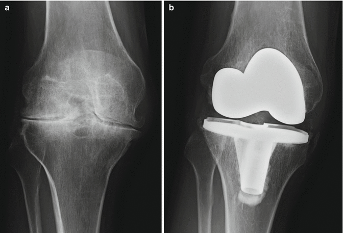
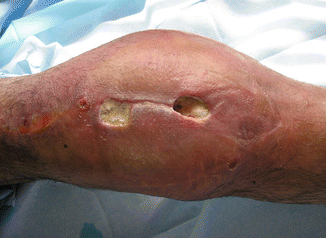
After 24–48 h, the drainage and intravenous antibiotic prophylaxis are removed, so that by the third day, the patient starts the postoperative rehabilitation. The patient is usually hospitalized for 7–10 days. The aim is for the patient to leave the hospital on foot with the help of walking sticks, with a knee flexion of 90° and total extension of the joint. The stitches or staples are usually removed after 2–3 weeks. The results of TKR in hemophilia have been quite satisfactory thus far (Fig. 8.2). Therefore, TKR is considered a good procedure in cases of severe knee arthropathy [5, 14–20]. The results are nearly comparable with those of osteoarthritis patients, and it can even be performed on patients with inhibitor [4]. However, the risk of infection is higher in hemophiliac patients than in the population with degenerative arthritis (osteoarthritis): average 7 % versus average 1 % (Fig. 8.3).


Fig. 8.2
Severe arthropathy of the knee (a) resolved satisfactorily with an unconstrained primary total knee replacement (TKR) (b)


