Histoplasmosis
Robert J. Leggiadro
INTRODUCTION
Histoplasmosis, caused by the dimorphic soil fungus Histoplasma capsulatum, is the most common pulmonary and systemic mycosis in humans. Normal as well as immunocompromised hosts may be affected, and reactivation disease is believed to occur during immunocompromised states.
EPIDEMIOLOGY
Although it is distributed throughout temperate zones of the world, histoplasmosis is recognized to be most highly endemic in the central United States. Factors known to favor fungal growth in soil are relatively high humidity, temperatures ranging between 20°C and 30°C, and strict aerobic conditions and acidity, which are characteristics of the highly endemic river basins of the central United States.
Outbreaks of symptomatic histoplasmosis are associated with point sources, or microfoci, of infection. Such microfoci are almost always characterized by contamination with bird or bat manure and include bird roosts, bat caves, old buildings, urban parks, decayed trees or wood piles, and chicken coops. Activities associated with H. capsulatum exposure and acquisition through the respiratory route include clearing or cleaning bird roosts and chicken coops, spelunking, construction or excavation, wood gathering or cutting, camping, and gardening. Dusty conditions greatly increase the number of infective airborne spores.
PATHOGENESIS
Human infection with H. capsulatum generally results from inhalation of airborne spores. These spores germinate into yeast forms within alveoli and bronchioles in 3 to 5 days. After an initial neutrophilic response, macrophages parasitize the yeast forms that proliferate within these cells in nonimmune individuals, eventually resulting in cellular death. Infected macrophages may also effect subclinical dissemination through the lymphatic system to regional lymph nodes, the liver, or the spleen. After a period of 1 to 2 weeks, specific cell-mediated immunity develops, which contains the infection and leads to resolution. Healing occurs slowly. Necrotic and caseous pulmonary and lymphatic lesions become encapsulated and calcified over months (children) and years (adults). Although they may persist in the central areas of caseous or calcified lesions, yeast forms are unlikely to cause further active infection. However, such endogenous foci may explain the occurrence of active histoplasmosis in immunocompromised individuals during residence in a nonendemic area. Presumably, these patients acquired histoplasmosis while residing in an endemic area before their immunosuppression. A possible alternative explanation to endogenous reactivation in such situations is exogenous reinfection from H. capsulatum microfoci in the environment of a nonendemic area.
CLINICAL MANIFESTATIONS
Most infections are asymptomatic or self-limited and do not require antifungal therapy. However, treatment is indicated in
disseminated and some other forms of H. capsulatum infection. Clinical features of acute pulmonary histoplasmosis, the most common form of self-limited infection, include fever, cough, chest pain, and fatigue. Chest radiographic findings range from focal infiltrates to hilar or mediastinal adenopathy and, rarely, a miliary pattern after intense exposure (Fig. 213.1). In the absence of treatment, symptoms improve gradually after several weeks. Nearly 10% of these patients develop either pericarditis or arthritis with erythema nodosum. Antiinflammatory, not antifungal, therapy is indicated for these individuals.
disseminated and some other forms of H. capsulatum infection. Clinical features of acute pulmonary histoplasmosis, the most common form of self-limited infection, include fever, cough, chest pain, and fatigue. Chest radiographic findings range from focal infiltrates to hilar or mediastinal adenopathy and, rarely, a miliary pattern after intense exposure (Fig. 213.1). In the absence of treatment, symptoms improve gradually after several weeks. Nearly 10% of these patients develop either pericarditis or arthritis with erythema nodosum. Antiinflammatory, not antifungal, therapy is indicated for these individuals.
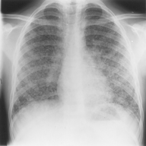 FIGURE 213.1. Miliary pulmonary histoplasmosis in an immunologically normal 10-year-old boy from Arkansas. There is a bilateral, diffuse, miliary, pulmonary parenchymal pattern of densities with greater involvement of the lower lobes. No hilar adenopathy or pleural effusion is present. (Reprinted from Robertson KR, James DH, Chesney PJ, et al. Miliary pulmonary histoplasmosis in a normal child. Arch Pediatr Adolesc Med 1994,148:833.) |
Antifungal therapy is indicated for patients with chronic pulmonary histoplasmosis, which is manifested by productive cough, sweats, weight loss, fatigue, and fibrotic apical pulmonary infiltrates with cavitation.
Fibrosing mediastinitis is a progressive and often fatal form of histoplasmosis that does not respond to either antifungal or antiinflammatory therapy. This excessive fibrotic reaction to past infection may improve after surgical resection of fibrotic mediastinal masses that are impinging on vital structures, although operative mortality is high.
Disseminated histoplasmosis may be seen in otherwise normal, healthy infants living in endemic areas or in immunocompromised individuals, most commonly those infected with the human immunodeficiency virus who are residing in, or have a history of residing in, an endemic area. These patients most frequently present with fever, hepatosplenomegaly, and pancytopenia. Chest radiographic features range from normal to focal or diffuse miliary infiltrates and adenopathy.
Related posts:

Stay updated, free articles. Join our Telegram channel

Full access? Get Clinical Tree


