9
Herpetic Whitlow
Kevin D. Plancher
History and Clinical Presentation
A 23-year-old nursing student, working in the intensive care unit for the first time, treated a patient without gloves. The patient reported symptoms of pain and burning or tingling of the infected digit. Erythema and edema followed with the development of vesicles on an erythematous base over the next 7 to 10 days. These vesicles are filled with clear or cloudy fluid.
Physical Examination
On examination, the patient’s finger is tender and edematous. Unlike a felon, the pulp space is not swollen. Examination revealed grouped vesicular lesions, which progressed to ulcers at 2 weeks (Fig. 9–1), and extension of the infection into subungual space and lymphangitic streaking was found. In the following 7 to 10 days the vesicles dried and began to heal (Fig. 9–2, different patient).
Diagnostic Studies
Diagnosis of herpetic whitlow is usually based on clinical presentation. The diagnosis can be confirmed with a Tzanck smear, which reveals characteristic multinucleated giant cells. Other smears, stains, and serologic tests can be used for diagnosis of primary infections. Herpes antibody titers can also be used to confirm the diagnosis.
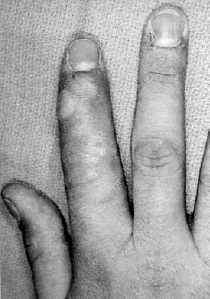
Figure 9–1. Grouped vesicular lesions of the index finger.
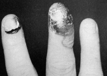
Figure 9–2. In the following 7 to 10 days the vesicles dry and begin to heal.
Related posts:

Stay updated, free articles. Join our Telegram channel

Full access? Get Clinical Tree


