Essentials of Diagnosis
- The sine qua non of Henoch-Schönlein purpura (HSP) is nonthrombocytopenic purpura, caused by inflammation in blood vessels of the superficial dermis.
- The pathologic hallmarks of HSP are a leukocytoclastic vasculitis and deposition of immunoglobulin (Ig) A in the walls of involved blood vessels.
- The tetrad of purpura, arthritis, glomerulonephritis, and abdominal pain is often observed. However, all four elements are not required for the diagnosis.
- More than 90% of cases occur in children. The disease is self-limited most of the time, resolving within a few weeks. Adult cases are sometimes more recalcitrant.
- Renal insufficiency develops in less than 5% of patients with HSP. The long-term renal prognosis depends mainly on the degree of initial damage to the kidney.
- HSP can be mimicked by other forms of systemic vasculitis that are more often life-threatening. For example, antineutrophil cytoplasmic antibody (ANCA)–associated vasculitides such as granulomatosis with polyangiitis (formerly Wegener granulomatosis) and microscopic polyangiitis (see Chapters 32 and 33, respectively) may also present with purpura, arthritis, and renal inflammation. Both of these disorders have the potential for serious involvement of other organs (eg, the lungs and peripheral nerves) and carry more dire renal prognoses.
General Considerations
Henoch-Schönlein purpura (HSP) is the most common form of systemic vasculitis in children, with an annual incidence of 140 cases per million persons. The peak incidence is in the first and second decades of life (90% of patients are younger than 10 years of age), with a male to female ratio of 2:1. The incidence is significantly lower in adults, with a mean age at presentation of 50 years. Males and females are affected equally and although HSP affects all ethnic groups, it is reportedly less common among blacks. Some epidemiologic studies suggest that HSP is more prevalent in the winter months.
HSP may be misdiagnosed as another form of vasculitis—most commonly hypersensitivity vasculitis (see Chapter 37)—because of the frequent failure to perform direct immunofluorescence testing on skin biopsy specimens. In two thirds of the cases, the disease follows an upper respiratory tract infection, with onset an average of 10 days after the start of respiratory symptoms. Despite this association, no single microorganism or environmental exposure has been confirmed as an important cause of HSP. HSP can also be induced by medications, particularly antibiotics. The American College of Rheumatology 1990 criteria for the classification of HSP are shown in Table 39–1. The first Chapel Hill Consensus Conference on the nomenclature of vasculitides defined HSP as a form of vasculitis characterized by the following: (1) IgA-dominant immune deposits within vessel walls; (2) small-vessel involvement (ie, capillaries, venules, or arterioles); and (3) skin, gut, renal, and joint manifestation.
|
The skin histopathology of HSP shows a leukocytoclastic vasculitis of small blood vessels within the superficial dermis. Necrosis is often present, but features of granulomatous inflammation are not. Immunofluorescent staining of biopsy specimens shows coarse, granular IgA staining in and around small blood vessels. In the kidney, the renal inflammation is indistinguishable from IgA nephropathy. There is a predilection for IgA deposition within the mesangium. However, in HSP nephritis, capillary wall staining for IgA is more frequently found and may be even more prominent than IgA in the mesangium. Most patients have increased serum IgA levels and circulating immune complexes that contain IgA, as well as IgA deposition in inflamed blood vessels.
Clinical Findings
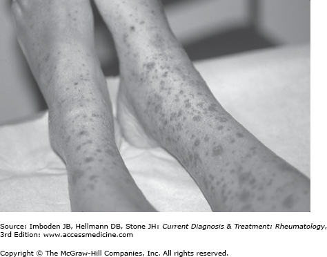
The classic full presentation includes the acute onset of fever, palpable purpura on the lower extremities (Figure 39–1) and buttocks, abdominal pain, arthritis, and hematuria. All components of this presentation are not required for the diagnosis, however. Conversely, even classic presentations are not diagnostic of this disorder. In adults, the diagnosis should be confirmed in most cases by biopsy (direct immunofluorescence as well as conventional hematoxylin and eosin staining). Pediatricians are more likely to rely on clinical diagnoses in the setting of classic presentations, which is reasonable given the relatively high incidence of HSP in children compared to adults.
The cutaneous findings of HSP include purpura (usually palpable, although sometimes not), urticarial papules, and plaques. Among adults, 60% of the patients have bullous or necrotic lesions (Figure 39–2
Related posts:


Stay updated, free articles. Join our Telegram channel

Full access? Get Clinical Tree


