Hematological treatment
Rest and splinting
Ice
Analgesia
Arthrocentesis (joint aspiration)
Arterial embolization
3.2.1 Hematological Treatment
On-demand therapy with a plasma-derived or recombinant FVIII or FIX concentrate is the first-line treatment for acute bleeding episodes in patients with hemophilia [1, 9]. Dosing ranges from approximately 20 to 40 IU/kg administered until bleeding stops. That means the infusion of FVIII 40 IU/kg at the time of joint hemorrhage and 20 IU/kg at 24 and 72 h after the first dose. Patients must be then encouraged to continue infusions of 20 IU/kg every other day, until joint pain and impairment of mobility had completely resolved.
Ultrasonography (USG) is very important in acute hemarthroses as it can be used to objectively identify the presence of blood in the joints, measure its size, assess its evolution, and confirm its complete disappearance [12].
In patients with inhibitors, rapid control of bleeding episodes using bypassing agents has the potential to minimize joint damage and to improve quality of life [13]. Smejkal et al. evaluated the efficacy and consumption of FEIBA in treatment of hemorrhages in hemophiliacs with factor VIII inhibitor [14]. The median cumulative dose of FEIBA per bleeding episode was 205 U kg (−1). Bleeding was stopped in 97 % of events, but re-bleeding occurred in 5 % of events within 48 h after cessation of bleeding.
A study indicated that frequently bleeding inhibitor patients are prescribed and use higher rFVIIa dosing for all bleed types than recommended in the package insert (90 mcg kg(−1)) [15]. The rFVIIa dosing was highly variable, particularly in the first days of treatment.
A global, prospective, randomized, double-blinded, active-controlled, dose-escalation trial evaluated and compared one to three doses of vatreptacog alfa at 5, 10, 20, 40, and 80 lg kg(−1) with one to three doses of rFVIIa at 90 lg kg(−1) in the treatment of acute joint bleeds in hemophilia patients with inhibitors [16]. A high efficacy rate of vatreptacog alfa in controlling acute joint bleeds was observed; 98 % of bleeds were controlled within 9 h of the initial dose in a combined evaluation of 20–80 lg kg(−1) vatreptacog alfa.
After administration of the appropriate doses of factor concentrates, pain will rapidly diminish, although inflammation and limitation of articular mobility commonly disappear more slowly. The degree of inflammation and limitation of motion are always related to the amount of blood in the joint.
3.2.2 Rest and Splinting
Rest for lower limb bleeding episodes should include bed rest (1 day) followed by avoidance of weight bearing and the use of crutches when ambulating and elevation when sitting (3–4 days).
For the knee a compressive bandage is adequate, although in very painful cases the bandage should be supplemented with a long-leg posterior plaster splint. For the ankle, a short-leg posterior plaster splint is recommended. For the upper limb, usually a sling (for the shoulder) or a long-arm posterior plaster splint (for the elbow) will provide sufficient rest, support, and protection. Lifting and carrying heavy items should be avoided until the bleeding has resolved (4–5 days).
3.2.3 Ice
Ice therapy could help to relieve pain and reduce the extent of bleeding, although its current role in hemophilia remains controversial. Experimental cooling of blood and/or tissue can significantly impair coagulation and prolong bleeding. In persons with hemophilia with acute hemarthrosis, ice application could impair coagulation and hemostasis [17].
3.2.4 Analgesia
For pain, paracetamol should be administered. Usually the hematological treatment provides adequate relief. Aspirin-containing products must be avoided. Unfortunately, there are no detailed algorithms or guidelines for pain management in hemophilia patients [18].
3.2.5 Joint Aspiration (Arthrocentesis)
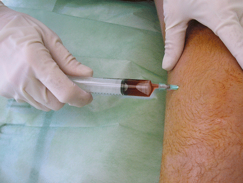
Joint aspiration is not commonly performed, but in cases of severe bleeding, it may relieve the patient’s pain and speed up rehabilitation (Fig. 3.1). There is a great deal of controversy on the role of arthrocentesis in hemophilia.


Fig. 3.1
Arthrocentesis (joint aspiration) in acute hemarthrosis of the knee in a hemophiliac. This must be done to relieve the pain and avoid the risk of future joint damage, although always with suitable hemostatic cover
Arthrocentesis should be performed in major hemarthrosis (very tense and painful joints) [19]. Joint aspiration should always be done under factor coverage and in aseptic conditions, in order to avoid recurrence or septic arthritis. When hemarthrosis does not respond to hematological treatment, septic arthritis must be suspected, especially if the patient is immunodepressed; joint aspiration and culture will allow us to reach a diagnosis [20].
If hemarthrosis does not respond to hematological treatment, one must suspect hemophilic synovitis, which can be detected by clinical examination. USG and magnetic resonance imaging (MRI) will help confirm the occurrence of synovitis. In such cases only aggressive treatment of synovitis will allow us to control articular bleeding, which is secondary to synovial hypertrophy. Synovitis can be controlled with early prophylactic treatment or by synovectomy (radiosynovectomy or arthroscopic synovectomy). Diagnostic imaging is paramount to assess the response to any type treatment.
Heim et al. [21] reported an interesting case of a person with hemophilia who had a fixed flexed hip and intractable pain. This clinical picture was suggestive of hemorrhage in that area. USG confirmed the diagnosis of acute hip hemarthrosis. Narcotic drugs failed to alleviate the severe pain. Joint aspiration produced dramatic pain relief and early joint rehabilitation. However, Heim et al. did not suggest that every coxhemarthrosis should be aspirated. It should be remembered that raised intra-articular pressure may contribute to femoral head necrosis in adults or to Perthes’ disease in children.
It is important to emphasize that while arthrocentesis of the elbow, knee, and ankle are quite simple procedures that can be done at the outpatient clinic, both shoulder and hip joint aspirations require sedation and radiographic control by an image intensifier, that is to say, they are surgical procedures done in an operating room, with an anesthetic and by an orthopedic surgeon.
3.2.6 Arterial Embolization
Klamroth et al. [22] reported seven patients who experienced recurrent massive bleeds that required arterial embolization. Under low-dose prophylactic treatment (15 IU fVIII or fIX per kg bodyweight for three times per week), no recurrent severe bleed unresponsive to coagulation factor replacement occurred after a mean observation time of 16 months after embolization. The consumption of factor concentrate decreased to one-third of the amount consumed before embolization.
In 21 cases of massive joint bleeding in 18 patients with hemophilia, selective catheterization was performed, and the bleeding was completely controlled by a single procedure in 14 cases [23]. Recurrence of the bleeding occurred in 7 cases and required a second embolization procedure; in one patient even a third embolization was required to stop the bleeding completely.
Selective angiographic embolization of the knee and elbow arteries was successful in 29–30 procedures [24]. Three patients remained free of bleeding events for more than 6 months. Additionally, after the procedure there was a significant reduction in factor FVIII usage that sustained up to 12 months after the procedures. No serious adverse events were observed. Therefore, angiographic embolization might be considered as a promising therapeutic and coagulation factor saving option in joint bleeds not responding to replacement of coagulation factor to normal levels [25].
Diagnosis and treatment of intra-articular hemorrhages must be delivered as early as possible. Additionally, treatment should ideally be administered intensively (enhanced on-demand treatment) until the resolution of symptoms. Joint aspiration plays an important role in acute and profuse hemarthroses. USG is an appropriate diagnostic technique to assess the evolution of acute hemarthrosis in hemophilia [26].
3.3 Recurrent Hemarthroses
Recurrent hemarthroses commonly occur after two or three articular bleeding episodes and persist despite adequate hematological treatment. Pain can be tolerable and is commonly associated with hypertrophic synovium on palpation and a slight lack of joint mobility.
When subacute hemarthroses recur for months and years, they will result in a state of hemophilic arthropathy. This usually occurs in young adults, who complain of persistent pain in the affected joint, not only with movement but also at rest. They may also have intermittent episodes of acute pain and inflammation related to synovitis or articular bleeding.
It is advisable to treat recurrent hemarthroses with hematological substitutive therapy, with two to three weeks of immobilization by means of a semiflexible splint. Some studies recommend 6–8 weeks of prophylaxis with physiotherapy. It is recommended to administer enough of deficient factor, three times a week, to obtain 20–30 % of the normal level. After each transfusion the patient should complete an exercise program focusing on active joint mobility, under the surveillance of an expert physiotherapist. If such mobility exercises are painful, only isometric exercises should be done.
When a flexion contracture does appear, it should be treated early and aggressively by conservative means to avoid it from becoming irreversible. Conservative measures include inverted dynamic splints, hinged extension–desubluxation casts, dynamic splints, and traction followed by a polypropylene orthosis. Inverted dynamic splints were specially designed for the knee joint and require admitting the patient to hospital. The lower limb is put in a balanced traction on a semicircular Thomas splint which has a knee flexion Pearson’s device. Then soft traction is put on the calf with the heel free; a posterior force is applied on the thigh by means of a cushioned spring located on the distal part of the thigh, which is connected by means of a string to a 3 kg weight. Such a posterior force counteracts the anterior force produced by the springs located on the posterior part of the calf. Both the longitudinal traction and the thigh weight are progressively increased. When the knee becomes fully extended, or if the technique does not work after one week of treatment, the patient is mobilized with a Böhler cast which is open in its anterior part.
The hinged extension–desubluxation cast can be made of plaster of Paris or of a thermoplastic material; it should be open in its anterior part. The hinge is adjusted once or twice a day to correct the deformity. When the contracture is less than 20°, the cast can be removed and replaced with a plaster splint. Hematological substitutive therapy is necessary during the procedure. The dynamic splint is adjustable and allows a low intensity but long duration force through the knee joint. A gain of 5–10° of knee extension can be expected in 6–9 months with this procedure. However, many patients may have hemarthrosis during the follow-up. Traction followed by orthosis is another alternative.
Stay updated, free articles. Join our Telegram channel

Full access? Get Clinical Tree


