Gymnastics
Larry Nassar
Female artistic gymnastics is one of the most popular summer Olympic sports in the United States. Tickets are in high demand to attend the competition, and millions watch the television broadcast during the Olympics. Yet during the quadrennium between Olympics, it is almost forgotten by the general public. In the medical community it is thought of as a sport of young girls, but this has changed. The Federation of International Gymnastics has been increasing the age of participation at the Olympics. Currently, the minimum age to be eligible for the Olympics is 16, and in the United States there are several international elite gymnasts who are over 20 years old and one that is 30 years old.
Gymnastics is a sport in constant flux. Every 4 years the rules change. With the rule changes come more difficult requirements. The equipment has dramatically changed over the years and a completely new vault table has recently been introduced. These frequent changes make it difficult for the general public to understand the sport and challenge the sports medicine practitioner to understand the current injury epidemiology.
EPIDEMIOLOGY
The most common areas for injury in the club and collegiate female gymnast (1) in order of frequency are (a) the ankle, (b) the wrist, and (c) the spine. Brüggemann, the current President of the Scientific Commission of the Federation of International Gymnastics, recorded that at takeoff for gymnastic tumbling on a spring floor (roundoff to a back-tuck somersault), the resultant moments at the ankle joint were 310 N·m in the sagittal plane and 100 N·m in the frontal plane (2). This correlates to an Achilles tendon force of 7,500 N (˜15 times the gymnast’s body weight), a tibiotalar joint force of 11,000 N (˜23 times body weight) and a talonavicular joint force of 8,000 N (˜15 times body weight). These forces in the foot and ankle are almost doubled with improper foot eversion or pronation by decreasing the contact area of the joint surfaces. With these types of extreme forces, the likelihood of injury of the foot and ankle is high.
LOW BACK INJURIES
Current elite-level female gymnasts are reported to have an 11.1% incidence of spondylolysis and a 2.9% incidence of spondylolisthesis (2). These statistics do not significantly differ from the normal population, yet a 44.4% incidence of osteochondritic changes was found in female gymnasts. The majority of these changes were end-plate compression fractures, where the nucleus pulposus herniates into the vertebral body. In the immature spine, especially during the adolescent growth spurt, the disc is stronger than the cartilaginous end plate. Thus, the end plate becomes damaged before the disc when exposed to compressive forces. These fractures are mainly found in the thoracolumbar junction (T11-L3). This area of the spine is at high risk for fracture due to its relative neutral alignment and transition between the more rigid thoracic spine and more flexible lumbar spine (1).
Swärd et al. found that normal disc heights were present with first diagnosis of vertebral end-plate damage in the gymnast. However, subsequent follow-up revealed marked disc
degeneration 10 to 12 months after injury (3). Nassar and co-workers studied the 1999 USA Gymnastics Female Artistic National Team, and 11 of 19 gymnasts (58%) had lumbar disc degeneration on magnetic resonance imaging (MRI). Anterior apophyseal ring fractures were found in 7 of 19 gymnasts (37%) (4).
degeneration 10 to 12 months after injury (3). Nassar and co-workers studied the 1999 USA Gymnastics Female Artistic National Team, and 11 of 19 gymnasts (58%) had lumbar disc degeneration on magnetic resonance imaging (MRI). Anterior apophyseal ring fractures were found in 7 of 19 gymnasts (37%) (4).
The skills and equipment in gymnastics have changed over the years, so findings in older studies on gymnasts are outdated and of little value. In the past, gymnasts performed many back walkovers, front walkovers, limbers, and other skills that created exceptional stress to the lumbothoracic spine. These skills are no longer performed with the same frequency as in the past. Gymnastic coaches have improved their technique with emphasis on proper use of the shoulders and thoracic spine to remove stress from the lower lumbar pars interarticularis. The addition of new training aids such as sting mats can reduce tumbling impact forces on the spine by 20% (2). Finally, reduction in the incidence of posterior column fractures may be attributed to coaches’ and athletes’ awareness to seek medical help early on for these injuries when only a stress reaction may be present and before a full stress fracture occurs.
The high-impact landing forces in gymnastics can be tolerated by the gymnast only by appropriate timing and balance of the muscular activity of the spine and lower extremity to distribute these forces most efficiently through the spine. If the gymnast performs a routine with improper timing or muscular firing sequence, the potential for injury on landing becomes great. Landing with a flexed trunk increases the pressure on the anterior end plates most significantly at the thoracolumbar junction. This leads to end-plate damage and marked disc degeneration noted 10 to 12 months after injury (3).
Hall states that the main cause of low back pain in the female artistic gymnast is uncontrollable hyperextension of the lumbar spine (5). Brady showed that a decrease in hip extension and poor thoracic extension motion contribute significantly to low back pain (6). Jull and Janda coined the term pelvic crossed syndrome (7) to explain how a gradual-onset nontraumatic overuse injury may develop in accordance with the explanations of gymnast lumbar mechanics given by Hall and Brady.
Pelvic crossed syndrome is an imbalance in the lumbar-pelvic-hip complex whereby the short and tight hip flexors and lumbar erector spinae muscles neurologically inhibit the proper firing and strength of the abdominal and gluteal muscles (7). This arrangement enhances an anterior pelvic tilt, increases lumbar lordosis, and shortens the hip flexors. The thoracolumbar erector spinae and hamstrings compensate by increasing activity, which creates hip extension.
The syndrome continues with gluteus medius weakness, which causes compensatory increased activity in the ipsilateral iliotibial band, tensor fasciae latae, and quadratus lumborum. The abdominal muscles display weakness with increased iliopsoas muscle activity, which increases trunk flexion. Sherrington’s law of reciprocal innervation has been proposed to explain this muscle inhibition. This type of muscle imbalance is self-sustaining without appropriate therapeutic intervention (8). When the inhibited muscle is exercised to create a maximal contraction, the electromyographic activity of the muscle actually decreases. No matter how specific the method of strengthening is, the muscle imbalance pattern is only reinforced unless the hypertonic/shortened muscle is first stretched (8).
MANUAL MEDICINE APPROACH
Principles in Gymnastics
Effective manipulations on gymnasts are a customized combination of techniques. The clinician should prepare his or her manipulations as a master gourmet chef would prepare a special meal for his guests. The clinician should combine the manual medicine techniques together in just the right order to best fit the overall condition of the athlete.
In general, it is recommended that the clinician perform articulatory and joint play techniques as described by Mennell for the examination and treatment of joint dysfunction in the extremities of the gymnast (9). By ensuring that the joints of the extremities are functioning at their highest capacity, the clinician may add
improved quality of movement for the athlete. Hypermobile joints in gymnasts should not be treated with articulatory, high-velocity, low-amplitude (HVLA) techniques mainly because they attempt stability with hyperlaxity.
improved quality of movement for the athlete. Hypermobile joints in gymnasts should not be treated with articulatory, high-velocity, low-amplitude (HVLA) techniques mainly because they attempt stability with hyperlaxity.
Regions
Two important body regions to focus on in gymnasts are the foot/ankle and the hand/wrist. The foot has chronic dysfunctions in the first metatarsophalangeal joint, cuboid, and tarsal navicular, while the ankle typically requires treatment in the subtalar joint and the proximal tibiofibular joint. For the wrist, close evaluation of wrist flexion-extension and supination-pronation motions of the forearm is recommended. The scapulothoracic articulation must be evaluated in conjunction with the foot and wrist, following along the kinetic chain principles.
Caution is warranted at the glenohumeral and scapholunate articulations, since these areas tend to be hypermobile. Research is needed to accurately detail the common areas of hyper- or hypomobility in the extremities of the gymnast.
Assessment and Application
The clinician who treats a gymnast during a competition needs to ascertain if the injury is acute or chronic, stable or unstable. It is not uncommon for a gymnast to arrive at a competition with a significant sprain, strain, or fracture for which she has not received any prior medical evaluation or treatment. The gymnast is generally a strong athlete with a very high pain threshold, and her gym performance is not always an accurate indicator of injury severity. An extreme case occurred at a junior Olympics national championship, when an adolescent gymnast sustained a fall onto her head 2 weeks before the competition. She continued to train and came to the meet hoping for manipulative treatment and clearance to compete. Her symptoms were severe enough that she did not compete and radiographs determined that she had a fracturedislocation in her cervical spine requiring surgical stabilization.
Because of this loose correlation between injury and perceived pain, articulatory or HVLA techniques should be avoided in acute injury unless proper imaging studies are available. Attempting to mobilize an unstable hypermobile segment of the body is also contraindicated. A rigid, spasmodic area should be approached cautiously because it usually involves the athlete’s splinting a fractured or strained area.
Length of treatment time also plays a factor in the selected techniques. During a competition, the gymnast competes and then rotates to the next event. In some cases the physician may have only 5 to 10 minutes to treat the gymnast between events. He or she may have only a chair and the floor to use as surfaces for treatment. The treatment may also be taking place in front of an audience and the media. Furthermore, the psychological makeup of the female gymnast needs to be understood. Is a doctor-patient relationship already established with the gymnast? Has the gymnast ever been manipulated before? Is she apprehensive to have someone treat her in a hands-on method? Thus, your selection of manipulative treatments must not only be appropriate for the injury, but also for the treatment setting and the psychological makeup of the athlete.
Myofascial Techniques
Soft tissue, articulatory, functional, muscle energy, myofascial release (MFR), HVLA, and craniosacral techniques are all appropriate treatments for the gymnast. These techniques should be used with the female gymnast as the clinician feels appropriate. The purpose of this chapter is to enlighten the reader on techniques employed frequently on the female artistic gymnast that may vary from typical treatments.
The obvious difference between gymnasts and other athletes is the need for extreme flexibility of the body while maintaining exceptional strength, power, and kinesthetic awareness. Manual medicine techniques are meant to maximize the gymnast’s range of motion, restore normal physiologic function, and yet not enhance pathologic ligamentous instability.
The pelvis is the center of the body’s link between the trunk, the upper extremity, and the lower extremity. It acts as an anchor for many vital tendons, ligaments, and fascial attachments.
Treatments to the pelvic girdle and its attachments can significantly benefit the gymnast by increasing mobility of the trunk, the lower extremity, and even the upper extremity without enhancing pathologic hypermobility. The pelvis and its attachments are used as the principal anatomic site treated in this chapter.
Treatments to the pelvic girdle and its attachments can significantly benefit the gymnast by increasing mobility of the trunk, the lower extremity, and even the upper extremity without enhancing pathologic hypermobility. The pelvis and its attachments are used as the principal anatomic site treated in this chapter.
Myofascial Release-Combination Technique
Rationale:
This is a combination of sustained longitudinal pressure MFR with longitudinal soft tissue stroking and digital ischemic pressure coupled with oscillating vibrations. This allows the connective tissue to elongate without exaggerating any ligamentous instability. Myofascial release spans the spectrum of manual medicine procedures and combines many of the principles of soft tissue, muscle energy, functional, and craniosacral techniques. MFR-combo techniques can be applied to the iliotibial band, paraspinal iliopsoas, ilioinguinal, and hamstring muscles, which are common sites of dysfunction in gymnasts.
Basic Technique
The treatment region is prepared with a small amount of massage lotion to prevent skin irritation.
The clinician applies perpendicular pressure to the contour of the area to be treated with his or her forearm. The amount of pressure is in general light to firm, about 10 to 15 lb. Do not push hard enough to bruise the tissue or cause undue pain.
The general path to follow is from distal to proximal; the speed of movement is about 1 in. per second.
The clinician repeats the process three to five times or until he or she feels the tissue tension resolve. If tender/trigger points or fascial restrictions are encountered, stop and apply more direct, longitudinally oriented pressure to the area with the elbow.
In addition, the clinician can rotate his or her forearm briskly (supinate and pronate) to send an oscillating vibratory force into the area, which helps to release the area more quickly and with less discomfort than with direct pressure alone. This type of myofascial release variation can be performed with the hand, forearm, or elbow. With this technique, deep slow breathing is an effective myofascial enhancer.
Variation: Gluteal Myofascial Release Technique
The gymnast lies prone on the treatment table.
Using the MFR-combo technique, the gluteal region is prepared with a small amount of massage lotion, and the clinician applies perpendicular pressure to the contour of the gluteal muscle with the forearm (Fig. 32.1).
The clinician starts at the thigh-gluteal junction and progresses through the entire gluteal region, ensuring that all of the hip internal and external rotators are completely treated. The process is repeated three to five times or until he or she feels the tissue tension resolve.
The gluteus medius is the most common muscle with symptomatic tender points in the gymnast, far more than the piriformis. Travell considers the gluteus medius to be the lumbago muscle because it is so intimately involved with low back pain (11).
Tender Point Release for Gluteus Medius
Rationale:
This technique uses ischemic digital pressure combined with a variation of strain-counterstrain and myofascial release. The technique was developed to meet the specific needs of the gymnast. Travell’s gluteus medius trigger points correlate closely to Jones’s points at the lower pole of the fifth lumbar vertebra, high ilium-sacroiliac joint, and third and fourth lumbar vertebrae (11,12). On occasion, the hip is extended as described by Jones if the foldover alone does not relieve the pain completely. Releasing the sacrotuberous ligament is a vital part of treating the gymnast discussed later in the chapter. Rarely, trigger point injections are needed (11).
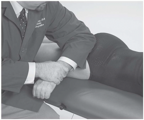 FIGURE 32.1. Gluteal myofascial release technique. |
The gymnast is prone with the clinician sitting caudad to the region being treated.
The tender point is located, ischemic digital pressure is applied, then the clinician folds the gluteal muscle bulk over the tender point in a cephalad-lateral rotation-type motion. This foldover should help resolve or greatly reduce pain from the digital pressure (Fig. 32.2).
The position is held along with the digital pressure for 30 to 90 seconds or until the clinician feels the tissue release. Jones recommends removing the digital pressure while holding the counterstrain position; however, with this type of treatment the digital ischemic pressure is maintained.
The gymnast should be instructed to take deep, slow breaths to assist with the release.
The digital pressure over the tender point and the foldover of the gluteal muscle should be returned toward neutral slowly so as not to precipitate the dysfunction from returning, as Jones recommends.
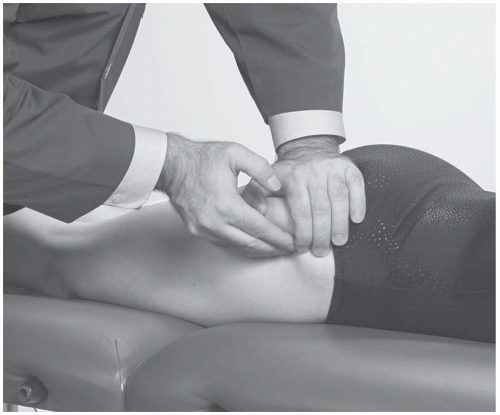 FIGURE 32.2. Gluteus medius tender point release. |
INJURY ASSESSMENT
Optimal function of the pelvis is vital to allow for the extreme range of motion, power, and balance needed to perform artistic gymnastics. The pelvis is the major connection between the upper and lower extremities; therefore, the pelvis should be evaluated for dysfunction with a large variety of injuries, such as ankle, shin, quadriceps, hip flexor, hip adductor, iliotibial band, gluteal region, pelvic bones, abdominal musculature, spine, and shoulder complex.
Sacrotuberous Ligament
The sacrotuberous ligament forms a functional bridge between the ankle and the shoulder, extending from the ischial tuberosity to the coccyx, and fans out toward the posterior sacroiliac joint capsule and the posterior superior iliac spine. The proximal tendon of the long head of the biceps femoris frequently attaches to the sacrotuberous ligament, functionally connecting it to the ankle (14). The biceps femoris tendon distally attaches to the fibula and the investing fascia of the peroneus longus.
In addition, the gluteus maximus, piriformis, multifidus, and thoracolumbar fascia all have direct attachments to the sacrotuberous ligament (15). The sacrotuberous ligament is directly connected to the upper extremity and upper trunk by the following two pathways:
The erector spinae aponeurosis and the iliocostalis thoracis.
The gluteus maximus is connected to the lumbodorsal fascia and linked to the contralateral latissimus dorsi (10).Related posts:

Stay updated, free articles. Join our Telegram channel

Full access? Get Clinical Tree


