Genitourinary Injury


Genitourinary Injury


James J. Thomas

Richard A. Santucci

Allen F. Morey


Although rarely lethal in nature, genitourinary tract injuries may be associated with significant morbidity. Prompt recognition and treatment are imperative to reduce complications and improve outcomes. Hematuria is a common but imperfect indicator of urinary tract injury. Because of ongoing treatment requirements for nonurologic associated injuries, radiographic evaluation of the urogenital tract must be efficient and appropriately conducted, while erroneous or unhelpful negative studies must be avoided. Although minor injuries can be safely observed, maximal urinary drainage and/or immediate reconstruction are often appropriate means of management in more severe injuries. Prompt surgical reconstruction of most genital injuries usually leads to adequate and acceptable cosmetic and functional results. In this chapter, we review the contemporary evaluation and treatment of genitourinary injuries in the trauma patient.


RENAL INJURIES


Evaluation


Presentation

Blunt rapid deceleration injuries are the most common etiology for renal trauma. Although blunt renal injuries tend to be minor in nature, some may be associated with damage to the renal vessels, renal artery thrombosis, or renal pedicle avulsion. Major renal injuries are often associated with multiple associated nonurologic injuries in the setting of rapid deceleration or penetrating trauma.

Hematuria is the best indicator of urinary system injury, although the degree of hematuria and the severity of the renal injury may not correlate. Microscopic hematuria seems to herald the presence of renal injury more commonly when shock is present,1,2,3 and less so when it is not. Microscopy is not necessarily required to evaluate hematuria, as the dipstick method is rapid and has a sensitivity and specificity for detection of microhematuria of more than 97%.4


Indications for Renal Imaging

The indications for radiographic evaluation after blunt trauma are gross hematuria, microscopic hematuria with shock, and major deceleration injury in the presence of significant nonurologic-associated injuries.3,5,6 Blunt trauma patients with isolated microhematuria do not in general require immediate imaging. Penetrating injuries with any degree of hematuria should be imaged.

Pediatric patients sustaining blunt abdominal trauma are at greater risk for renal injury than adults because children have less perirenal fat to protect and stabilize the kidney.1 Shock may be a less useful criterion in children to determine if imaging studies should be performed.7 Children with insignificant microhematuria (<50 red blood cell [RBC] per high power field [HPF]) after blunt trauma are a low-yield group and usually do not require urgent renal imaging.7


Imaging Studies

Computed tomography (CT) is the gold standard for the radiographic assessment of stable patients with renal trauma and should be performed whenever possible in cases where renal injury is suspected.5,8,9 CT will accurately determine the location and depth of renal lacerations, the presence and amount of urinary or vascular contrast extravasation, as well as preexisting renal abnormalities. The American Association for the Surgery of Trauma (AAST) organ injury severity scale is the preferred classification scheme for grading of renal injuries, classified by abdominal CT or direct renal exploration (see Table 1).6


TABLE 1 AMERICAN ASSOCIATION FOR THE SURGERY OF TRAUMA (AAST) ORGAN INJURY SEVERITY SCALE FOR THE KIDNEY


Grade


Description of Injury


1


Contusion or nonexpanding subcapsular hematoma


No laceration


2


Nonexpanding perirenal hematoma


Cortical laceration <1 cm deep without extravasation


3


Cortical laceration >1 cm without urinary extravasation


4


Laceration: through corticomedullary junction into collecting system


or


Vascular: segmental renal artery or vein injury with contained hematoma, or partial vessel laceration or vessel thrombosis


5


Laceration: shattered kidney


or


Vascular: renal pedicle or avulsion


(Adapted from Moore EE, Shackford SR, Pachter HL, et al. Organ injury scaling: spleen, liver, and kidney. J Trauma. 1989;29(12): 1664-1666.; Nicolaisen GS, McAninch JW, Marshall GA, et al. Renal trauma: Re-evaluation of the indications for radiographic assessment. J Urol. 1985;133(2):183-187.)


Failure of the kidney to opacify after contrast administration is a hallmark of renal pedicle injury. Central parahilar hematoma is suggestive of renal pedicle injury, even if the renal parenchyma is well enhanced. In all cases of suspected renal trauma evaluated with spiral CT, additional delayed scans should be performed 10 to 15 minutes after contrast injection to evaluate the integrity of the collecting system.10

Unstable patients selected for immediate surgical intervention (and therefore unable to have a CT scan) should undergo one-shot intravenous pyelogram (IVP) in the operating suite if they need evaluation of renal or ureteral injury. The technique consists of a bolus intravenous injection of 2 mL per kg radiographic contrast followed by a single plain film taken after 10 minutes. The study is safe, efficient, and of high quality in most cases.11 It provides important information concerning the injured kidney, and confirms a normal functioning kidney on the contralateral side. Although ultrasonography is a popular imaging modality in the initial evaluation of abdominal trauma, its role in staging renal trauma is not well established at this time.12


Treatment


Nonoperative Management

The kidney has remarkable healing properties, and nonoperative management has therefore become the treatment of choice for the vast majority of non-life-threatening renal injuries. Nonoperative management results in an excellent long-term outcome in most cases. All grade 1 and 2, and most grade 3 renal injuries can be managed nonoperatively. Exploration of grade 4 and 5 renal injuries often results in nephrectomy; recent data indicates that many of these patients can be managed safely with an expectant approach.13,14

Patients with suspected penetrating renal injury who are otherwise stable should undergo radiographic staging, whenever possible, to define the injury. Renal gunshot injuries require exploration only if they involve the hilum or are accompanied by signs of continued bleeding, ureteral injuries, or renal pelvis lacerations.15 Stab wounds and low-velocity gunshot wounds may often be managed conservatively with an acceptably good outcome.16 Tissue damage from high-velocity gunshot injuries may be more extensive.

The site of penetration by stab wound has an important influence on management—if posterior to the anterior axillary line, most (88%)17 may be managed nonoperatively.18,19 A systematic approach based on clinical, laboratory, and radiologic evaluation may minimize negative exploration after renal stabbing, without increasing morbidity from missed injury. Expectant management of renal stab wounds can be attempted on the hemodynamically stable patient, especially if ureteral and renal pelvis injuries can be ruled out. Ultimately, 98% of blunt renal injuries can be managed nonoperatively.

Grade IV and V injuries more often require surgical exploration, but even many of these can be managed without renal surgery if carefully staged and selected. Patients with high-grade injuries selected for nonoperative management should be closely monitored for persistent bleeding with repeat vital signs and serial hematocrits. If significant urinary extravasation persists beyond 48 hours, prompt urologic consultation with retrograde placement of an internal ureteral “double J” stent (and a urethral catheter to prevent urinary reflux) will often prevent prolonged urinary extravasation and improve perirenal urinoma formation, sepsis, and ileus.20 Follow-up abdominal CT scans are reserved for high-grade injuries and symptomatic patients only (dropping hematocrit, flank pain, fever, etc.). Should renal bleeding persist, or delayed bleeding occur, angiographic studies with embolization of bleeding vessels will often obviate surgical intervention. It is popularly believed that attempts to repair a bleeding kidney in the days after injury when inflammation is maximal (say 3 to 30 days) will inevitably result in nephrectomy, so it seems prudent to attempt expedient angioembolization if possible.


Angiographic Techniques

Angiography is helpful in defining the exact location and degree of vascular injuries and is useful when planning selective embolization. Arteriography with selective renal embolization for hemorrhage control is a reasonable alternative to laparotomy, provided no other indication for immediate surgery exists.21 The rate of successful hemostasis by embolization is reportedly identical in blunt and penetrating injuries.22,23


Operative Management: Indications

The only absolute indication for renal exploration is life-threatening hemodynamic instability due to renal hemorrhage, irrespective of the mode of injury.24 Another strong indication for renal exploration is an expanding or pulsatile perirenal hematoma identified at exploratory laparotomy (this finding heralds a grade 5 vascular injury and is quite rare). Relative indications for surgery include suspected renal pelvis injury or persistent bleeding (≥3 units per 24 hours). It has been recognized that most injuries that have urinary extravasation and devitalized kidney fragments heal with nonoperative treatment.25


Renal Reconstruction

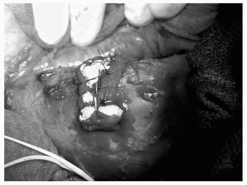
Renal reconstruction (renorrhaphy) by debriding, oversewing, and covering the defect is feasible in most cases (see Table 2), but partial nephrectomy may be required when large amounts of tissue are damaged, especially at the pole of the kidney. Obtaining early vascular control before opening the Gerota fascia can decrease renal loss caused by bleeding during attempted renal repair (see Fig. 1).26 In a series of 133 renal units in which early vessel isolation and control before opening the Gerota fascia was achieved, McAninch et al.24 reported a very high renal salvage rate of 89%. Early vascular control does not increase postoperative azotemia or mortality.


Renovascular Injuries

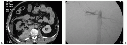
Renovascular injuries are associated with extensive associated trauma and increased perioperative and postoperative mortality and morbidity. Prompt nephrectomy is usually the treatment of choice except in those very rare cases in which there is a solitary kidney or the patient has sustained bilateral vessel injuries (see Fig. 2).17 Attempts at surgical repair of renal artery thrombosis largely fail, and certainly after 8 hours the kidney cannot be salvaged. Many patients with renal vascular injury are critically injured, with numerous associated organ injuries, low body temperature, and poor coagulation; the advisability of major vascular repair over a nephrectomy is limited in the unstable patient if a normal contralateral kidney is present. Damage control with placement of packs and planned return for corrective surgery within 24 hours is another reasonable alternative option.27 Because the kidney is a paired organ it is sometimes sacrificed with alacrity in patients who would not truly be in danger from an attempted repair. In a recent series of 1,360 adult patients with renal lacerations 23% underwent surgery, and an appalling 64% of these got a nephrectomy.28 This harmful practice must be avoided.


TABLE 2 PRINCIPLES OF RENAL RECONSTRUCTION AFTER TRAUMA


Consider preliminary vascular control—occlude vessels with Rommel tourniquet if major injury


Complete renal exposure


Judicious debridement of nonviable tissue


Hemostasis by individual suture ligation of bleeding vessels (4-0 chromic on RB-1 needle)


Watertight closure of the collecting system (either by suturing or by closing overlying parenchyma)


Coverage or approximation of the parenchymal defect


Consider use of hemostatic agent such as fibrin sealant over repair


Perirenal drain separated from repair, placed through dependant incision


RB, round body.


Figure 1 Repair of midpole renal laceration with multiple capsule sutures tied over Gelfoam bolster.


Complications

Early complications of renal injury include bleeding, infection, perinephric abscess, sepsis, urinary fistula, hypertension, urinary extravasation, and urinoma. Delayed complications include bleeding, hydronephrosis, calculus formation, chronic pyelonephritis, hypertension, arteriovenous fistula, and pseudoaneurysm, although these are all less common. Urinary extravasation can usually be managed expectantly as most resolve spontaneously. In cases where there is persistent leakage with fever and/or sepsis, ureteral stent placement or percutaneous drainage is usually feasible and curative.


URETERAL INJURIES


Evaluation


Presentation

Unlike renal injuries, nearly all ureteral injuries occur as a result of penetrating or iatrogenic trauma. And unlike renal
injuries, the presence of hematuria is not a reliable indicator of ureteral injury. Many (25% to 45%) cases of ureteral injury will not have even microscopic hematuria,29,30 so a high index of suspicion is required.


Figure 2 A: Computed tomography (CT) showing poor contrast uptake into right renal artery, concerning for renal artery thrombus. B: Confirmatory angiogram shows abrupt cutoff of right renal artery consistent with thrombus.

The rare entity of ureteropelvic junction (UPJ) disruption consequent to rapid deceleration injury is associated with an unusual pattern of either medial or “circumrenal” contrast extravasation seen on CT.31 Children are felt to be more susceptible because of their hyperextensible vertebral column.32


Diagnosis

Wound location is felt to be the best indicator of ureteral injury, and direct exploration is the best means of detecting its presence. Intraoperative detection requires a high degree of suspicion. The trajectory of the knife/missile must be carefully examined and ureteral exploration undertaken in all cases of potential injury. Intravenous infusion or direct ureteral injection of indigo carmine will often produce a pool of colored urine, which may guide intraoperative detection of ureteral injury.

Liberal use of preoperative diagnostic tools such as CT is helpful, whenever possible. Vigilance for delayed presentation of ureteral injuries will also allow detection of injuries missed on presentation. Fever, leukocytosis, and local peritoneal irritation are the most common signs of missed ureteral injury and should always prompt CT examination.


Computed Tomography

Ureteral injuries often manifest in the absence of contrast in the ureter on delayed images. This underscores the absolute necessity of tracing both ureters throughout their entire course on trauma CT scans.33 Delayed images must be obtained 5 to 20 minutes after contrast injection.34 Medial extravasation of contrast or nonopacification of the ipsilateral ureter on CT remains the most reliable finding, especially in UPJ avulsion (see Fig. 3). A late finding of ureteral injury is a urinoma surrounding the ureter. A postcontrast kidney ureter and bladder (KUB) can supplement the CT images, which is created by getting a plain abdominal film after the CT with contrast is complete.


Retrograde Pyelography

Retrograde pyelography is used to delineate the level and extent of ureteral injury seen on CT scan or IVP, or if further clinical information is needed. At the same time, if appropriate, a retrograde ureteral stent can be attempted.


Treatment


Primary Repair

Repair of the ureter must be meticulous, and urologic consultation should be promptly sought to prevent adverse
medicolegal consequences. Ureteral blood supply is tenuous and the sequelae of imperfect repair include urine leakage, abscess, fistula, and refractory ureteral stricture, requiring additional surgical procedures and/or nephrectomy.


Figure 3 Delayed computed tomography (CT) scan with contrast showing medical extravasation of the right kidney, consistent with upper ureter or renal pelvis injury.

Primary repair is recommended in most ureteral injuries. Principles of primary ureteroureterostomy involve limited debridement followed by creation of a spatulated, watertight closure using optical magnification, with interrupted or running 5-0 or 6-0 absorbable monofilament such as Maxon (polyglyconate).29 An internal stent and retroperitoneal drain are placed in most cases. This technique is used primarily for injuries of the upper two thirds of the ureter.


Ureteroneocystotomy

Ureteroneocystotomy is used to repair ureteral injuries in the distal one third of the ureter. A tensionfree repair must be achieved—sometimes the ureteral stump can be reimplanted directly into the bladder. Other times, the bladder must be brought up to the ureter using either psoas hitch or Boari procedure, although complex reconstructions are often best reserved for a delayed setting (see Fig. 4). In trauma cases, we favor a simple refluxing, nontunneled anastomosis to decrease the chance of postoperative stenosis. Use of a stent or feeding tube across the anastomosis is advisable initially.

Only gold members can continue reading. Log In or Register to continue

Stay updated, free articles. Join our Telegram channel

Oct 17, 2016 | Posted by in MANUAL THERAPIST | Comments Off on Genitourinary Injury

Full access? Get Clinical Tree

Get Clinical Tree app for offline access