Fig. 67.1
Gaucher disease involving the humerus. Note the radiolucency with cortical scalloping involving the distal diaphysis; the trabecular pattern of the distal end is spared
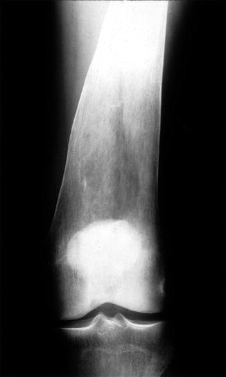
Fig. 67.2
Gaucher disease causing modeling deformity and cortical thinning of the femur. There is overall radiolucency of the femur, and the normal tapering cortical flare is widened, giving rise to the so-called Erlenmeyer flask deformity
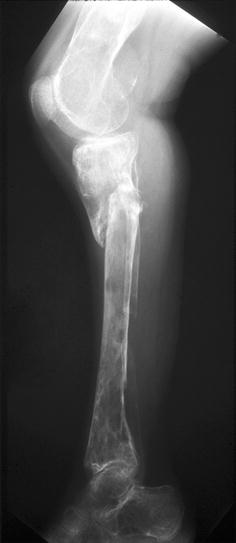
Fig. 67.3
Gaucher disease, leg. There are areas of radiodensity and space-occupying radiolucency secondary to deposits of Gaucher histiocytes with extreme attenuation of the tibial and fibular cortex leading to pathologic fracture. The distal femur demonstrates “bone within a bone” configuration suggesting old reactive periostitis
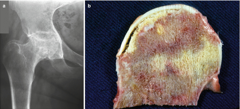
Fig. 67.4
(a) Gaucher disease with avascular necrosis. AP conventional radiograph of the right hip demonstrates a mixture of sclerosis and lysis of the superior femoral head with joint narrowing and deformity but without secondary osteophytes. This is consistent with stage 2 avascular necrosis with superficial collapse of the articular plate. (b) Gaucher disease with avascular necrosis. The resected gross specimen seen in (a) demonstrating irregular areas of necrosis and viable bone with separation of the subarticular bone plate and overlying articular cartilage, giving rise to the so-called crescent sign observed when this separation can be seen with routine radiographs
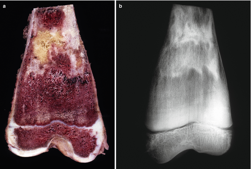
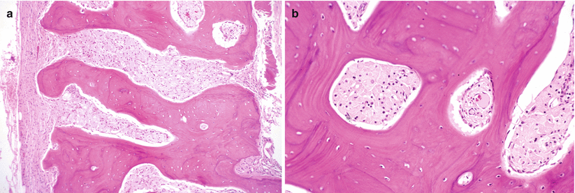
Fig. 67.5
(a) Gaucher disease, autopsy specimen of distal femur in a child unsuccessfully treated with enzyme therapy. The metaphyseal flare is convex rather than concave, accounting for the typical Erlenmeyer flask deformity, and the cortex is attenuated to almost paper thinness. The marrow, which is usually yellow at this site at this age, is red because of replacement of the more proximal marrow with Gaucher histiocyte storage. Note irregular whitish and yellow areas representing medullary infarctions. (b) Specimen radiograph of the femur seen in (a) demonstrates that the cortex is thinned almost to invisibility and the irregular radiodensities are secondary to dead bone and calcification of marrow fat in medullary fat necrosis (From Klein et al.; with permission)