Fig. 1.1
Facial appearance changes with age in a girl with FCMD; images were obtained at 7 months (A), at 1 year and 5 months (B), and at 8 years of age (C). Note that the myopathic face is not evident at 8 months (A). At 2 years, puffing of the cheeks and open mouth are evident (b). At 5 years, open mouth and a sharp chin are recognizable (c). A boy with FCMD at 1 year and at 3 years of age (D, E). He can sit on his own and is classified as having the typical form. Facial muscle involvement is not evident (D). He is training to maintain the standing position with HKAFO (E). (These images are shown with permission from the parents)
1.3 Diagnostic Testing
1.3.1 Serum Creatine Kinase (CK)
The level of serum creatine kinase (CK) is usually under 10,000 U/l, not as high as that of DMD patients. Under age 6 years, CK is 10–50 times higher than normal, but it decreases after age 6 years. CK decreases to the normal level when patients become bedridden. The level of CK changes depends on maximum motor development. In patients with the severe phenotype, who are bedridden, the CK level is highest in infancy or early childhood and then decreases linearly. In patients with the typical or the mild phenotype, the CK level increases with motor development, reaching the highest around the time of peak of their motor function, and then decreases with age and deterioration. Aspartate aminotransferase (AST), alanine aminotransferase (ALT), lactate dehydrogenase (LDH), and aldolase are 1.5–3 times higher than normal. FCMD patients are on occasion misdiagnosed as having liver dysfunction because ALT is sometimes higher than AST, a finding that also occurs in young children with DMD.
1.3.2 Neuroimaging
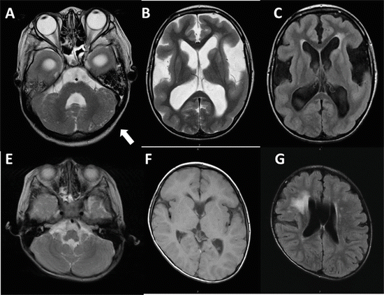
Typical findings of magnetic resonance imaging (MRI) in FCMD patients are cobblestone lissencephaly and cerebellar cystic lesions [14] (Fig. 1.2). Cerebral abnormalities on MRI are mainly classified into two patterns of cerebral cortical dysplasia. Both patterns are usually symmetrical and can be seen at all ages. The first pattern is typical of polymicrogyria showing a slightly thickened cortex with shallow sulci and a bumpy gray-white matter interface at the frontal lobe in all patients and at the temporoparietal lobes in some cases. Employing sagittal images may facilitate detecting the changes in this cortical dysplasia that were limited to the frontal lobe [14]. The second pattern is pachygyria showing a thick cortex with a smooth surface and a smooth gray-white matter interface in the temporo-occipital lobes. In the cerebellum, polymicrogyria such as disorganized cerebellar foliation with intermingled islands of granular and molecular layers and cerebellar cysts which are intraparenchymal cysts located in the peripheral hemispheres are typically seen in FCMD patients, regardless of age. White matter abnormalities with hyperintensity on T2-weighted images and hypointensity on T1-weighted images are seen especially in younger patients and those with severe phenotypes. Since the signal alteration varies in extent among patients and becomes milder with age, the issue of whether white matter abnormalities suggest delayed myelination, demyelination, dysmyelination, or other problems remains controversial [15–17]. Recent studies with MR spectroscopy indicate delayed myelination rather than dysmyelination [16, 17]. Dilated lateral ventricles are often recognized, and some patients with severe phenotypes may show hydrocephalus similar to that associated with WWS. A thin and rounded corpus callosum and cavities of the septum pellucidum or Berger’s space in older patients are often seen. Hypoplasia of the brain stem is sometimes seen, and sagittal views reveal a thin brain stem which is straighter than normal.


Fig. 1.2
Brain MR images in a 10-year-old girl with severe phenotype (A, B, C) and in a 3-year-old boy with typical phenotype (D, E, F). A T2-weighted image shows several small cysts (arrow) in the cerebellum (A). Pachygyria with a thickened cortex showing a smooth surface in the frontotemporal lobes can be seen. Wide Sylvian fissures and dilated ventricles are evident on the T2-weighted image. (B) The high-intensity area close to the frontal horn of the lateral ventricle in the white matter region was easily demonstrated on FLAIR (c). No apparent cysts were detected in the cerebellum on the T2-weighted image (D), and pachygyria in the frontal region was very mild on a T1-weighted image (E). High-intensity areas were also detected in a typical phenotype case employing FLAIR (F)
In Fig. 1.2, the three upper brain MRIs are from a severe-type patient, and the lower three are from a patient with typical-type FCMD. T2-weighted images show frontal pachygyria, dilated ventricles, and high myelin signals. The cerebellum has numerous pits. These abnormalities are more severe in the severe type.
1.3.3 Skeletal Muscle Imaging (MRI and CT)
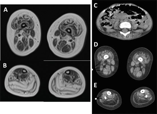
Skeletal muscle imaging detecting the patterns of affected muscles specific for each disease is useful for differential diagnosis or assessment of the progression of muscle disease. In FCMD patients, the changes with fatty replacement are more striking and extensive at the calf level (Fig. 1.3). The fatty changes in calf muscles in FCMD patients are already detectable in early infancy and progress rapidly as compared to those in DMD, indicating that this is one reason for FCMD patients never becoming ambulatory [18]. Muscle imaging showed marked involvement of the gastrocnemius and soleus muscles at the calf level, and the biceps femoris, vastus lateralis, and rectus muscles at the thigh level are also secondarily involved. Paraspinal muscles become involved at the early stage. On the other hand, the gracilis, sartorius, and tibialis posterior muscles are relatively preserved. Since there are patients with the mild phenotype who can become ambulant due to preserved soleus muscles, some extent of maximum motor development can be assumed based on skeletal muscle imaging.


Fig. 1.3
T1-weighted skeletal muscle MRI (A, B) and CT scans (C, D, E) in a 3-year-old FCMD patient. Reticulated increased intensities were recognized in the rectus femoris and vastus lateralis (A) and marked high-intensity areas in the soleus and gastrocnemius muscles. Atrophy and scattered low-density areas were seen in the paraspinal muscles at the T3 level (C) and low-density areas in the rectus femoris and vastus lateralis muscles, while the gracilis and sartorius muscles were well preserved (D). Low-density changes were seen throughout the soleus and gastrocnemius muscles (E)
1.3.4 Muscle Pathology
Findings such as necrosis, regenerating fibers, and increases in connective tissues are seen. Compared with those in DMD cases, all fibers are round and small. Some extremely small fibers (<10 μm) are scattered among other muscle fibers. There is no inflammatory cell infiltration. Hayashi et al. reported a selective deficiency of highly glycosylated α-DG, but not β-dystroglycan (DG), on the surface membranes of skeletal and cardiac muscle fibers in patients with FCMD [19]. Since the diagnosis can be made by genetic analysis and specific clinical features in most cases, muscle biopsy should not necessarily be performed for diagnostic purposes.
1.3.5 Neuropathology
The main central nervous system (CNS) lesion in FCMD patients is cobblestone lissencephaly of the cerebrum and cerebellum. Several studies investigating how fukutin works via α-DG in neurons have been conducted. The basement membrane covers the glia limitans where the glycosylated α-DG is observed. In CNS lesions of the fetus with FCMD, glioneuronal tissues protrude into the leptomeninges through disruption of the glia limitans in which the glycosylation of α-DG is reduced [20–22]. Hence, hypoglycosylation of α-dystroglycan is considered to be one of the main causes of glia limitans disruption, resulting in cobblestone lissencephaly. Astrocytes are also presumably involved in FCMD. As Hiroi et al. suggested, fukutin appears to be important not only simply for migration but also for cell differentiation. Their study demonstrated that the expression of FKTN mRNA decreased with maturation in the human cerebrum and raised the possibility that fukutin might be involved in cellular differentiation via glycosylation of α-dystroglycan. Immature neurons remain in an immature state during migration and begin to develop into a mature form once they reach the proper position after migration. Fukutin might be important for preventing inappropriate differentiation during migration [23].
1.4 Complications and Management Strategies
1.4.1 Intellectual Involvement
Mental retardation is seen in most FCMD patients, and the level is moderate to severe [24], with IQs ranging from 30 to 50 [25]. Most typical patients master very little speech, fewer than 20 meaningful words, and are never capable of sentence formation. The severity of mental retardation usually correlates with phenotypes classified by maximum motor development, that is, severe-form patients can learn no or only a few words, while those with the mild form can speak in sentences. Some patients with refractory epilepsy can learn only a few words though they are able to sit without support or slide on the buttocks, showing a discrepancy between speech ability and motor development. There are a few reports on the mild-form FCMD with a mild mental deficit. One report described a mild-form patient, heterozygous for a 3-kb insertion of the ancestral founder mutation, whose IQ on the Tanaka-Binet scale was 97 [26]. Since no mutation was detected in the coding region of the other allele in this patient, the phenotype-genotype correlation remained unclear. Social development is not as severely affected as mental abilities [27]. FCMD patients are shy but enjoy communicating with people and can have positive relationships with other children in kindergarten and primary school. Autistic features are rare in FCMD patients.
1.4.2 Seizures
Because of cortical dysplasia, more than 50 % of FCMD patients exhibit febrile seizures and/or epilepsy beyond 1 year of age [2]. Yoshioka and Higuchi reported seizures in 80 % of their FCMD patients, and the average age at onset was 3 years [28]. Usually, the initial seizures occur with a febrile episode, though one-third of patients had afebrile seizures from the onset [28]. Most patients show generalized tonic or tonic-clonic convulsions, as febrile seizures, early in life. Later, however, the main convulsive disorder becomes complex partial seizures. In most patients, epilepsy is mild and treatable, but some patients develop intractable seizures including Lennox-Gastaut syndrome. West syndrome has also been documented but is rather rare in FCMD patients [29, 30]. Yoshioka et al. reported the correlation between seizure severity and genotype and concluded that mutation analysis of the FKTN gene may predict seizure outcomes [30]. In their study, heterozygotes usually developed seizures earlier (the onsets of febrile and afebrile seizures were, on average, 3.6 and 3.7 years) than homozygotes (5.4 and 4.6 years), and some heterozygotes had intractable seizures.
In our recent study, 62 % of patients developed seizures. Among them, 71 % had only febrile seizures, 6 % had afebrile seizures from the onset, and 22 % developed afebrile seizures following febrile seizures. A comparison of seizure prevalences between homozygotes and heterozygotes showed a slightly higher rate in the latter, but the difference did not reach statistical significance. The peak age at febrile seizure onset was 1–2 years and the mean age was 3 years, later than that of febrile convulsions in the normal population. Mean age at afebrile seizure onset was 5.8 years, showing no peak. Most patients have seizures that are controllable with just one type of antiepileptic drug, but 18 % have intractable seizures that must be treated with three medications. Comparison between patients with seizures controllable with one type of antiepileptic drug and those requiring more than two types revealed no statistically significant differences in genotype or phenotype, but there was a tendency for the number of intractable seizures to be lower in homozygotes. There was also a tendency for the total number of seizures and intractable seizures to be lower in homozygotes. In contrast to a previous report, the author’s study group found that intractable seizures can manifest in homozygotes, especially those with severe intellectual involvement, despite relatively good motor development and mild abnormalities on brain MR images. Fixed discharges in the F areas tended to be a common feature in the intractable cases. Paroxysmal discharges on electroencephalography did not usually have a fixed location and were independent of the brain MRI abnormalities. Carbamazepine and valproic acid (VPA) are the agents most frequently used and show no statistically significant difference in efficacy. We have experienced a few FCMD patients treated for intractable epilepsy with many types of antiepileptic drugs, including relatively a high dose of VPA, with kidney tubule cell damage progressing to Fanconi syndrome. Instead of high-dose VPA, levetiracetam (LEV), which has recently been administered for intractable epilepsy as a second-line therapy in Japan, is now among the effective choices for seizures complicating FCMD. LEV is effective for both partial and generalized seizures, and we have succeeded in controlling intractable seizures with LEV in some of our FCMD patients. No serious side effects have been reported, and LEV is especially useful for patients with respiratory dysfunction and/or dysphagia since it does not increase oropharyngeal secretion, a common side effect of antiepileptic drugs.
1.4.3 Ocular Abnormalities
Ocular abnormalities including refractive errors and accommodation disorders are seen in almost half of FCMD patients. Retinal abnormalities are mild and focal, but the prevalence is relatively high, being present in 32 % of FCMD patients with the more severe phenotype [2, 31]. Refractive errors (myopia and hypermetropia) are common, and microphthalmia, retinal detachment, and cataracts are seen in patients with the severe phenotype [27]. Hino et al. demonstrated the clinicopathological features of the eyes of FCMD patients in detail [32]. Their histopathological examinations revealed various retinal abnormalities even in FCMD cases without ophthalmological alterations. The authors raised the possibility that the initial changes in retinal dysplasia may occur during the fetal period. Reductions in basement components, cytoskeletal proteins, and glutamate metabolism-related proteins produced by Müller cells in the eyes of FCMD patients implicate defective Müller cell properties in the retinal pathology of FCMD. Fukutin may be highly relevant to the cell-to-cell interaction of retinal blasts with Müller cells, and downregulation of FKTN mRNA may thus be responsible for retinal dysplasia [32]. Regular ophthalmologic evaluations are recommended especially for patients with the severe phenotype.
1.4.4 Respiratory Failure
Progressive respiratory failure typically emerges early in the second decade of life and is one of the most common causes of death in FCMD patients. Noninvasive positive pressure ventilation (NPPV) has been used as an effective therapy, although tracheostomy with invasive ventilation (TIV) may be needed in some cases. The author’s research group retrospectively studied chronic respiratory failure in genetically diagnosed FCMD patients. In our study, the mean age at the introduction of noninvasive positive pressure ventilation support was 12 years. NPPV is beneficial and well tolerated despite the intellectual impairments. A few patients received tracheostomy with invasive ventilation (TIV) after age 15 years without prior NPPV, because of difficulty with extubation followed by a respiratory infection and some urgently required laryngotracheal separation after episodes of aspiration pneumonia or suffocation. Heterozygotes tended to develop the complication of chronic respiratory failure earlier than homozygotes. Respiratory failure also progressed earlier in the severe-type patients. Though the difference did not reach statistical significance, chronic respiratory failure manifests earlier in those with poor motor function and/or a heterozygous genotype. It is often difficult at the outpatient clinic to detect respiratory dysfunction in FCMD patients. Most patients cannot perform lung function tests mainly due to mental retardation and because they are unable to purse their lips due to facial muscle weakness. Furthermore, the symptoms of chronic respiratory dysfunction such as sleep disturbance or poor intake are difficult to detect since many FCMD patients have had these symptoms since they were quite young. Regular assessments with end-tidal CO2 monitoring during sleep should be conducted once a year to detect respiratory dysfunction, starting at about age 10 years in typical-form patients and much earlier in those with the severe form.
1.4.5 Cardiomyopathy
Cardiac involvement usually develops very slowly, first appearing after 10 years of age, in FCMD patients. Since Hayashi et al. reported a deficiency of α-DG in the cardiac as well as in the skeletal muscles of FCMD patients [19], it is reasonable that cardiac involvement also manifests in both DMD and Becker muscular dystrophy (BMD). Fibrosis of the myocardium was confirmed at autopsy in several reports [33, 34], and moderate-to-severe multifocal fibrosis was observed throughout the left ventricular (LV) wall, especially in the anterior, lateral, and posterior walls [34]. The degree of fibrosis was greater in patients who died of heart failure than in those who died of other causes such as respiratory problems. Electrocardiographic (ECG) abnormalities are common and similar to those in DMD and BMD. Especially, a tall R wave on V1 with an R/S ratio >1 is observed in most FCMD patients, while deep but narrow Q waves on leads I, aVL, V5, and V6 are not seen as frequently as in dystrophinopathies. In contrast, cardiomegaly on chest radiography is rare. Plasma brain natriuretic peptide is not elevated till the advanced stage of cardiac dysfunction and is not useful for early detection. An echocardiogram is the most useful method of detecting the early stage of cardiac dysfunction. A significant correlation between age and LV fractional shortening (LVFS) was confirmed and LVFS decreased with age. LVSF was normal in most patients under 10 years of age but was reduced in most patients over 15 years of age. Therefore, a regular cardiac evaluation including echocardiograms and follow-up is important in patients aged 10 years and older. It is noteworthy that there was no significant difference in LVFS among the three phenotypes. Also, LV function in heterozygous patients is similar to that in younger children who are homozygous. There is no definite relationship between cardiac dysfunction and either the severity or the genotype of FCMD. Rather, we can say that the frequency of cardiac involvement tends to become higher in mild and typical FCMD cases. There is an involvement discrepancy between cardiac dysfunction and skeletal muscle systems, which is also seen in DMD or BMD. We hypothesized that the greater mobility of mild and typical cases may place more of a burden on the hearts. It is rare, but a few patients rapidly developed serious cardiac dysfunction and died before 15 years of age, and a few patients with the severe form showed LVFS reduction before 5 years. Once the presence of cardiac involvement is recognized, patients are usually treated with angiotensin-converting enzyme (ACE) inhibitors or a combination of ACE inhibitors and β-blockers. These interventions were reported to improve LV function in patients with DMD and BMD [35]. Combination therapy with ACE inhibitors and β-blockers for 2 years was also found to significantly increase LVFS, and this regimen was more efficient than therapy with only ACE inhibitors in several types of muscular dystrophies including FCMD [36].
Stay updated, free articles. Join our Telegram channel

Full access? Get Clinical Tree


