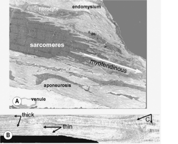
3.1 Force transmission and muscle mechanics There are two fundamental ways to look at force transmission. 1. The direct (mechanics) way: considers a force transmitted from the sarcomeres of a muscle onto, for example, the tendon and from there to bone to cause movement of a body segment. 2. The inverse mechanics way: deals with questions: which structures are the sources of so-called reaction forces that allow the muscle to exert force? An active or stretched passive muscle will shorten unless opposed by a load (an opposing force) that will prevent shortening to a length at which no force can be generated (slack length). This opposing force is a reaction force that is equal to the force exerted by the muscle (action = reaction), but has an opposite direction. It may be exerted by the tendon or by other structures arranged in series with the sarcomeres. Each myofiber (muscle fiber) is equipped (at least at one end) with a myotendinous junction (Fig. 3.1.1A). The thin filaments of the last sarcomere of myofibrils within myofibers are attached sideways through the sarcolemma to collagen fibers of the aponeurosis (tendon plate) that invade the invaginations of the myofiber but remain outside of the cell. The supramolecular structures involved in such connections are shown in Fig. 3.1.2A–C. The myotendinous loads exerted on the last sarcomere are transmitted to the next sarcomeres in series within each myofibril. If this is the only type of force transmission, forces in all sarcomeres in series within a myofibril need to be equal. If this is the exclusive reaction force available, and if all sarcomeres have identical properties, such sarcomeres will shorten until their identical force is in equilibrium with the reaction force. In static final conditions, this will yield identical sarcomere lengths within the myofiber. Fig. 3.1.1 • The myotendinous junction.
General principles
Myotendinous force transmission

(A) Low power electron micrograph showing this junction of one myofiber. The intracellular parts (e.g., sarcomeres) are dark, and light areas are extracellular materials. A considerable area of contact between the two exists because of many invaginations of the myofiber. The aponeurosis is made up from many little tendons belonging to single myofibers. (B) High power electron micrograph showing one invagination containing collagen fibrils (C) that will form the myofiber’s small tendon (further to the right). Within the intracellular part thick and thin filaments of the last sarcomere are indicated. Across the sarcolemma and basal lamina (dark line border of the invagination and myofiber) the thin filaments are connected to the collagen fibrils.![]()
Stay updated, free articles. Join our Telegram channel

Full access? Get Clinical Tree


Musculoskeletal Key
Fastest Musculoskeletal Insight Engine


