Joseph S. Park, Venkat Perumal
Foot and Ankle
Foot and Ankle
General Guidelines
Preoperative antibiotics are given half an hour before the incision is made
A tourniquet is applied according to the surgeon’s preference
An Esmarch bandage or pneumatic tourniquet may be used
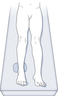
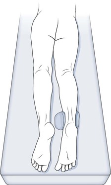
All bony prominences should be adequately padded
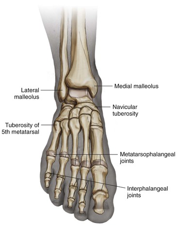
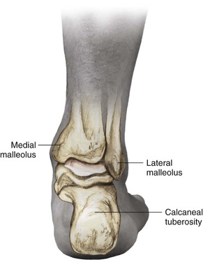
Review the pertinent anatomy and mark the palpable landmarks
Regional Anatomy
Osteology
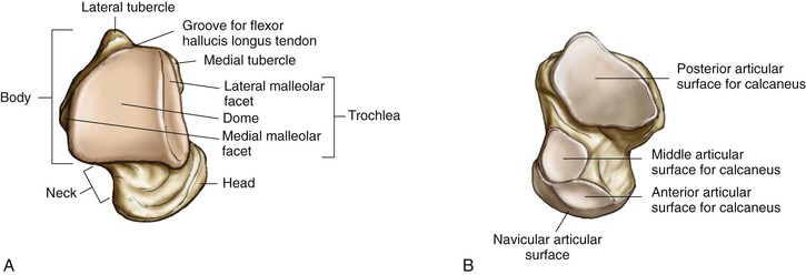
Talus (Fig. 8-1)
Three parts: body, neck, and head
No muscles or tendons are attached
The superior surface is a dome with a large articular facet
The lateral surface has a triangular lateral malleolar facet
The inferior surface has a large concave posterior facet for the calcaneus
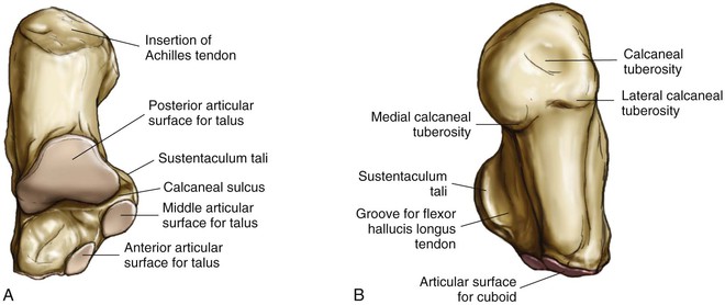
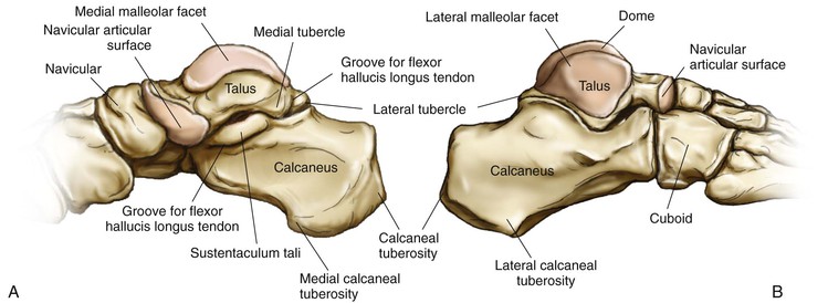
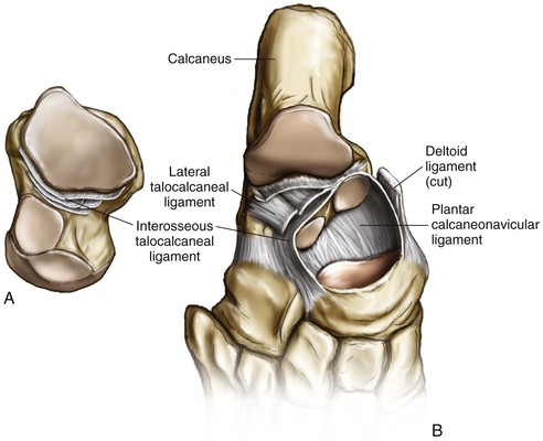
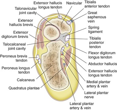
Calcaneus (Fig. 8-2)
The posterior surface has an area for insertion of the Achilles tendon
The anterior surface is triangular and concavoconvex and articulates with the cuboid
The groove of the calcaneus is between the posterior and middle facets, and it opens laterally to a rough quadrangle
The thick medial border of the sustentaculum, with the tendons of the tibialis posterior above and the flexor digitorum longus (FDL) on its medial margin, is grooved inferiorly by the tendon of the FHL
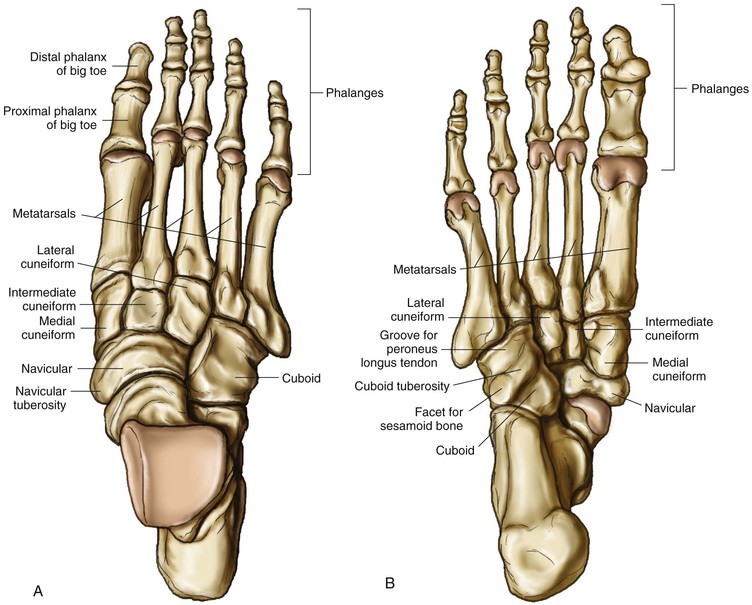
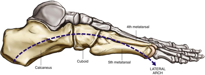
Cuboid (Fig. 8-3)
The anterior surface articulates with the fourth-fifth metatarsal base
The posterior surface articulates with the calcaneus
The superior surface has ligamentous attachments
The inferior surface has tuberosity and a groove for the peroneus tendon
The medial surface articulates with the late cuneiform and with the navicular
Sesamoids
The sesamoids are situated where tendons cross joints and change the direction of pull of tendons
The sesamoids also improve the mechanical advantage of the tendon
The medial and lateral sesamoids are constant
The following sesamoids are inconstant:
• Sesamoid of the tibialis posterior tendon
• Sesamoid in the peroneus longus tendon
• Sesamoids under the metatarsal heads, commonly under the second and fifth heads
Arthrology
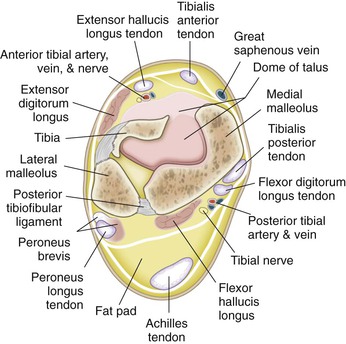
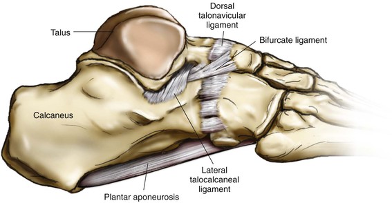
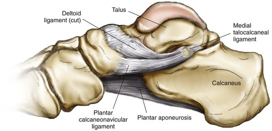
Ankle Joint (Figs. 8-5 to 8-7)
The ankle joint is a strong and stable joint
The ankle joint is a synovial hinge joint
A capsule encloses the joint and is attached to the bony articular margins
• Anterior talofibular ligament
• Posterior talofibular ligament
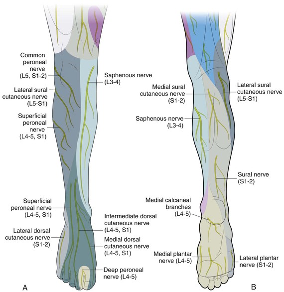
• Deep peroneal and tibial nerve
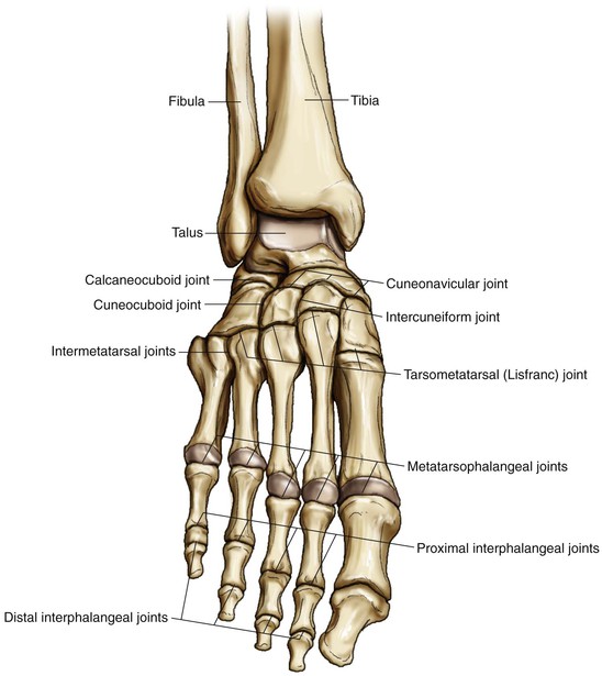
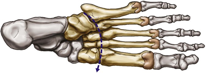
Metatarsophalangeal (MTP) and Interphalangeal Joints (see Fig. 8-8)
MTP joints are condyloid joints
Stabilized by plantar plates and oblique collateral ligaments
Deep transverse ligaments connect all five MTP joints
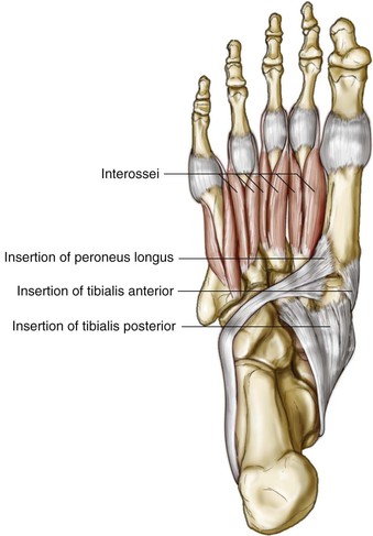
The second toe is long axis, and adduction-abduction is performed by interossei
Interphalangeal joints are hinge joints
• Interphalangeal joints only provide plantar flexion movement
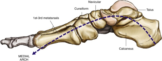
Arches of the Foot
Muscles
Extrinsic Muscles
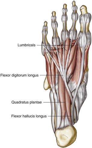
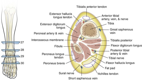
Muscles are in the leg, but their tendons function within the foot
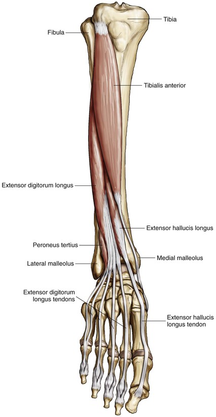
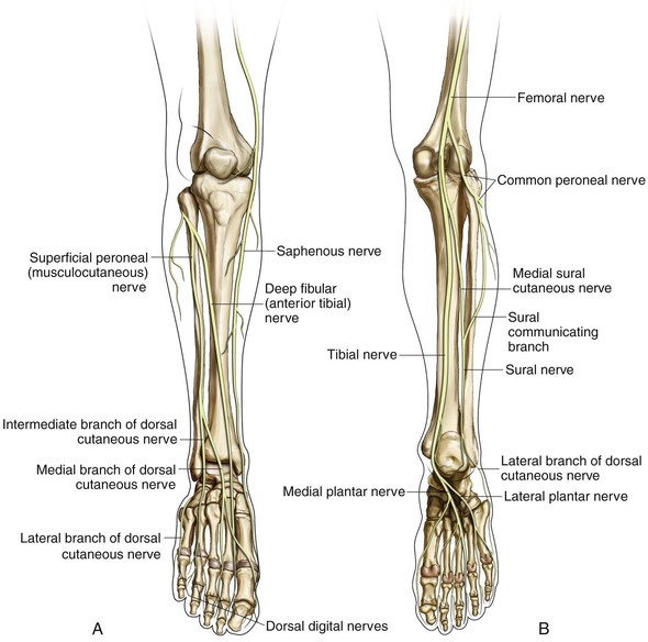
• Anterior compartment (innervated by the deep peroneal/anterior tibial nerve; Fig. 8-12)
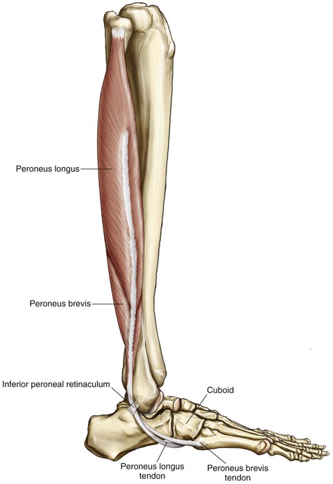
• Lateral compartment (innervated by the superficial peroneal nerve; Fig. 8-13)
• Peroneus longus and brevis muscle
• Tendons descend together in a common tendon sheath underneath the superior peroneal retinaculum
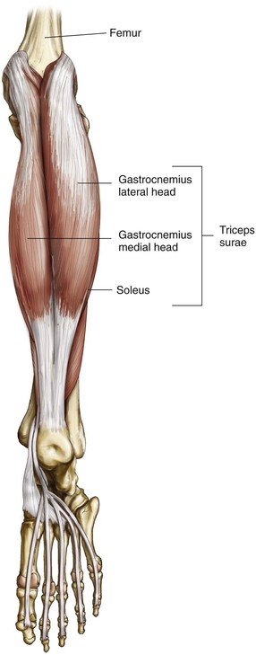
• Posterior compartment (innervated by the posterior tibial nerve; Figs. 8-14 to 8-16)
• Flexor digitorum longus (FDL)
• Flexor hallucis longus ( FHL)
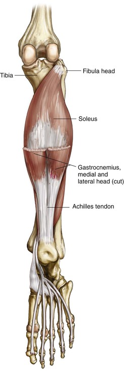
• Assisted by the peroneals, FHL, and FDL
• TA
• Assisted by the EHL, EDL, and peroneus tertius
• Eversion
• Peroneus longus, brevis, and tertius
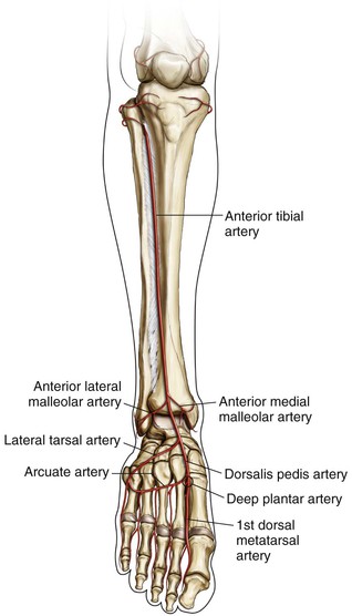
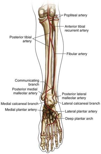
Vascularity (Figs. 8-23 and 8-24)
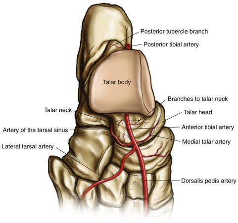
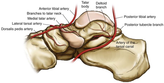
Blood Supply of the Talus (Figs. 8-25 and 8-26)
• Superior surface of the neck
• Medial talar arteries—medial recurrent tarsal artery (branch of the anterior tibial artery)
• Inferior surface of the neck
• Artery of the tarsal canal (branch of the posterior tibial artery)
• Deltoid branch (posterior tibial artery)
• Direct branch from the posterior tibial artery—infrequently from the peroneals
• Superior neck vessels and branches of the tarsal sinus artery
Surgical Approaches to the Ankle
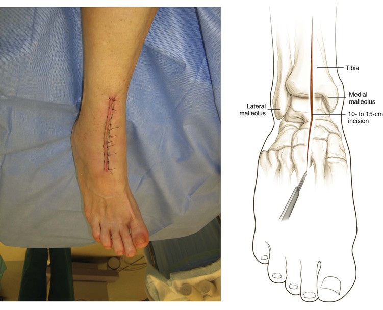
Anterior Approach to the Ankle (Video 8-1)
Indications
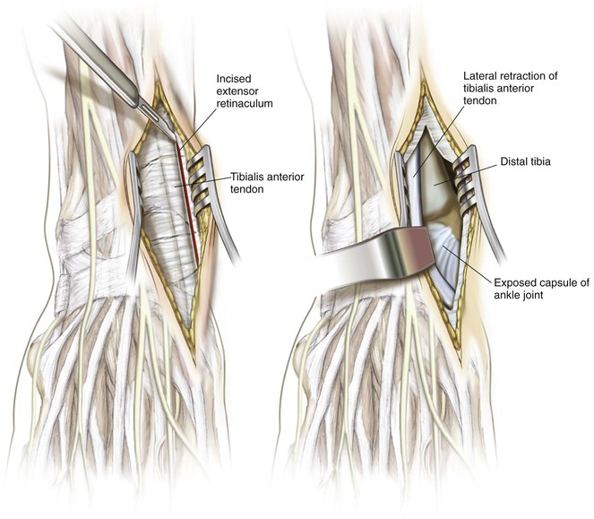
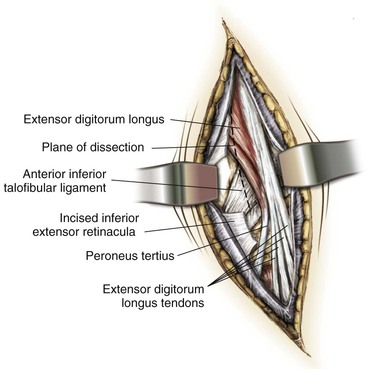
Superficial Dissection (TA/EHL Interval)
Superficial Dissection (Alternative—EHL/EDL Interval)
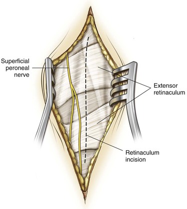
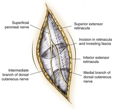
Incise the deep fascia of the leg in line with the skin incision
Incise the extensor retinaculum
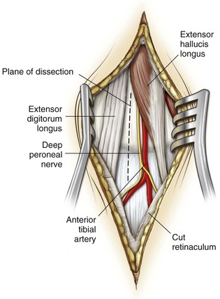
Find the plane between the EHL and EDL a few centimeters above the joint (Fig. 8-40)
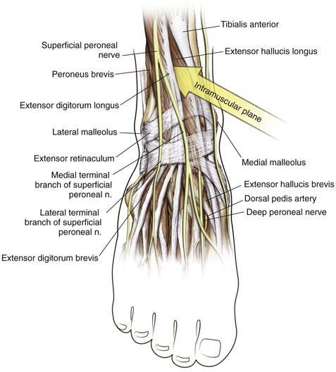
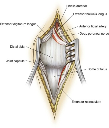
Identify and mobilize the anterior tibial artery and deep peroneal nerve
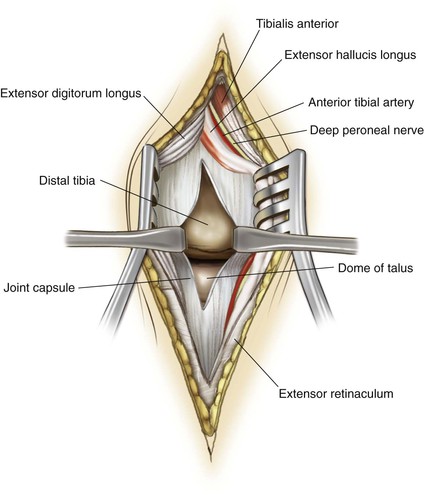
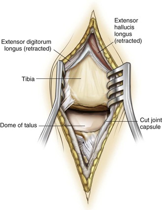
Retract the EHL and neurovascular bundle medially and the EDL laterally
This approach allows for distal extension to approach the dorsal mid foot (Fig. 8-41)
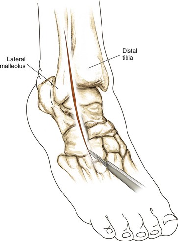
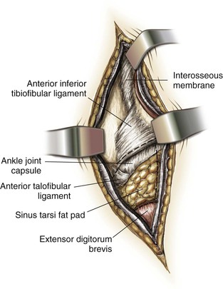
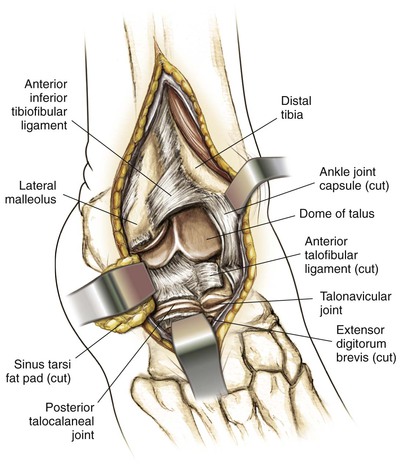
Anterolateral Approach to the Ankle
Indications
Direct Posterior Approach to the Achilles Tendon
Indications
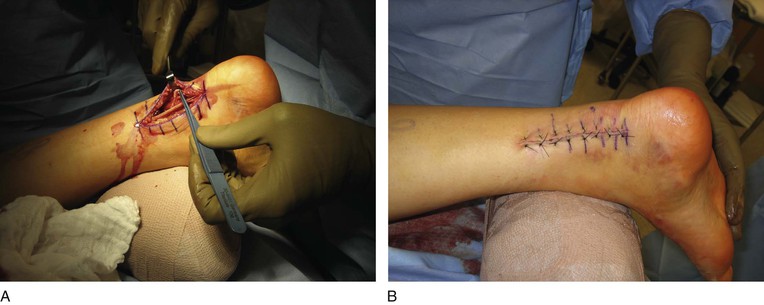
Surgical Exposure (Figs. 8-51 and 8-52)
For acute Achilles tendon ruptures
• A longitudinal incision is made 1 cm medial to the Achilles tendon to improve soft tissue coverage over the tendon after repair (Fig. 8-51); the deep fascia over the Achilles tendon is incised medial to the tendon
In an alternative approach an incision is made just lateral to the Achilles tendon (Fig. 8-52)
In patients with chronic tendinopathy, a midline posterior incision over the tendon is typically used to allow access to the calcaneus (Fig. 8-53)
• The paratenon is incised in the midline so that the paratenon can be closed after the reconstruction
Stay updated, free articles. Join our Telegram channel

Full access? Get Clinical Tree


