Chapter 8 Fixation of Osteochondral Fragments
Introduction
Osteochondral fragments can also be created by the surgeon in the course of performing osteochondral autografts such as mosaicplasty and mega OATS (Chapters 5 and 6) or osteochondral allografting (Chapter 7).
This chapter reviews the most common methods of fragment fixation.
Indications for Internal Fixation
Few studies provide clear indications for surgical fixation of osteochondral fragments (Table 8-1). The underlying diagnosis has implications in the decision-making process, such as in the treatment of osteochondritis dissecans (OCD).
TABLE 8-1 Indications for Internal Fixation with Regard to Type of Osteochondral Fragment
| Type of Osteochondral Fragment | Indication for Fixation |
|---|---|
| Osteochondral fracture Chondral fracture Osteochondritis Dissecans (OCD) | Fragment is nondeformed, reducible, viable Lesions that are unstable, partially detached lesions, completely detached lesions that are nondisplaced and viable displaced lesions |
To our knowledge, no quantitative measures of graft stability are available to assist the surgeon in deciding when to perform adjunctive fixation.
Juvenile Osteochondritis Dissecans
Surgical indication for juvenile osteochondritis dissecans (jOCD) includes failure of 6 to 9 months of nonoperative treatment of a stable lesion, an unstable or detached lesion, or a symptomatic patient with imminent physeal closure within 6 months to a year.1
Adult Osteochondritis Dissecans
Unlike jOCD, symptomatic adult OCD necessitates surgical intervention in most cases because of the progressive course, the inability of the fragment to heal on its own, and the fact that most adult lesions are unstable in nature.2
Massive lesions, multiple fragmented lesions, and substantial craters often require procedures utilizing open bone grafting of the bed or osteochondral autografts and allografts when fixation is not possible or the fragment is not adequate for fixation.3
Traumatic Osteochondral Fragments
In adults there is little evidence that purely chondral fragments have the ability to heal to bone. Nonetheless, surgeons may choose to attempt to repair very thin or small osteochondral fragments with the understanding that there is a significant risk of failure.4,5
One caveat of this approach (and, in fact, in treating any osteochondral fragment with internal fixation) is the avoidance iatrogenic injury to the joint by the fixation device should the fragment collapse or detach, leaving the fixation device proud or loose within the joint. Numerous case reports have documented articular cartilage injury and joint destruction by exposed or loose fixation devices.3–7
Overview of Internal Fixation Devices
The goal of any chosen device should be rigid fixation and compression while establishing a position that is seated low enough as to not interfere with the surrounding articular cartilage (Table 8-2). The device should allow for early range of motion and should retain the ability to be removed, if necessary.8
TABLE 8-2 Internal Fixation Devices
| Metal Pins and Wires |
| Smooth and threaded metal pins K-wires |
| Cannulated and AO Metal Screws |
| Constant pitch Variable pitch, headless (e.g., Herbert screw) |
| Bioabsorbable Implants |
| Biocompression screws Chondral pins and darts (smooth and barbed) |
| Osteochondral Plugs |
| Autograft bone plug/Osteochondral core |
Pins and Wires
The use of smooth metal pins for fixation was originally described by Smillie in 19579 but has since become more of a historical reference. Good results have been published using K-wires in combination with drilling and bone grafting,8,10 especially when splitting of a smaller fragment is a concern. Other advantages of K-wires include availability, low cost, and ease of use. Disadvantages include lack of compression achieved and possibility of breakage, along with the need for removal.
Cannulated and Variable Pitch Screws
Several options exist with metal screws including cannulated screws and variable-pitch, headless screws. The most commonly referenced variable-pitch screw is the Herbert screw.11–14 This type of screw allows for rigid fixation, as well as having an auto-compression effect. Another advantage to this screw is that the headless design allows it to be countersunk beneath the surface of the articular cartilage.
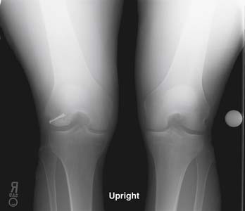
The disadvantage of the Herbert screw along with the other metal screws is that they may require an additional surgery for removal, and, if the fragment settles or breaks, the screw may become proud, leading to injury of the opposing articular surface. When possible, the surgeon attempts to place these devices in a manner that avoids the articular surface (Fig. 8-1).
< div class='tao-gold-member'>
Related posts:


Stay updated, free articles. Join our Telegram channel

Full access? Get Clinical Tree


