Fig. 5.1
Modelling of the cartilage destruction
5.1.2 Simulation of Loss of Carpal Height
The reduction of gaps has resulted in loss of carpal height. Carpometacarpal ratio (CR)—the ratio between the distance from the distal radius to the base of the third metacarpal with the length of the third metacarpal [4]—was utilised to simulate this condition. Further details were mentioned in Table 5.1 and the simulated loss of carpal height was shown in Fig. 5.2.
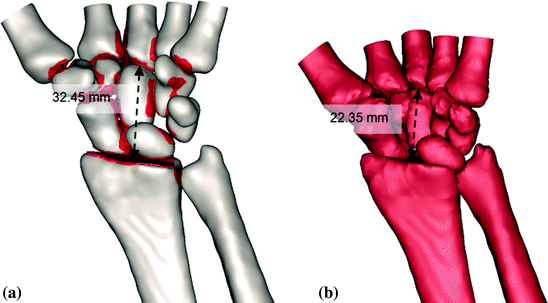
Table 5.1
Information on the carpometacarpal ratio used in this study in comparison with the literature
Literature [4] | Current study | Simulation |
|---|---|---|
Healthy: CR = 0.54 ± 0.03 | Healthy:  | Translation of metacarpals 10.1 mm proximally to simulate severe RA model |
Severe RA: CR = 0.40 | Severe RA: 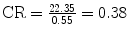 |
Fig. 5.2
Simulated loss of carpal height due to bone destruction (b). As compared to the healthy model (a) (total carpal height of 32.45 mm), the simulated impaction in RA model was seen to have total carpal height reduced to 22.35 mm
5.1.3 Simulation of Dislocation of the Carpus in the Ulnar Direction
Dislocation of the carpus in the ulnar direction occurs due to loss of tension of the radiotriquetral ligament, irrespective of the status of the ulnar head [2]. The entire carpus excluding the scaphoid was involved in the simulation. Figure 5.3a depicts the dislocated carpus towards ulnar. The simulation was done by rotating 10° of carpus towards ulnar with the center of the radius used as the center of rotation (COR). Figure 5.3b shows the simulated loss of tension (in circular) of the radiotriquetral ligament where only one link remained mimicking weakened ligaments.
Fig. 5.3
The dislocated carpus towards ulna (a). The RA bones are in red and the transparent bones represent the normal healthy wrist. Weakened ligaments were simulated by remaining one link (b). The ligament in circle was the simulated loss of tension of the radiotriquetral ligament
5.1.4 Simulation of Dislocation of the Proximal Carpal Row in the Palmar and Ulnar Directions
Dislocation of the proximal carpal row in the palmar occurred physiologically as during ulnar deviation (either due to physiological movements or as a results of disease), the scaphoid, lunate, and triquetrum rotate palmarly [16]. In the rheumatic wrist, the occurrence of SLAC (stage 3), SLD and the weakened radio-triquetrial ligaments has resulted in the dislocation of carpus towards ulnar direction [2]. These situations induced greater load subjected to the lunate thus ultimately destruct the capitolunate joint [5]. Figure 5.4 illustrates the simulated palmar (2.76 mm) and ulnar dislocation (7.61 mm) of the proximal carpal bones.
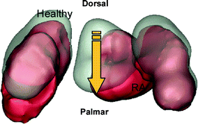
Fig. 5.4
Superior view of the proximal row carpal bones. The figure shows its dislocation towards palmar and ulnar directions. The RA bones were in red and transparent bones represent the normal healthy bones
5.1.5 Simulation of Scapholunate Dissociation and Scapholunate Advanced Collapse
The SLD was simulated by increasing distance between the scaphoid and the lunate, from 1.98 to 6.51 mm. The worn and torn intrinsic ligaments as results from the synovitis effect [2, 5] were simulated by utilising one link. As stated by Trieb et al. [2], the scapholunar and lunotriquetral ligaments are commonly effected as the disease progresses, thus this condition subsequently destruct the scapholunate articulation [9, 17, 18]. This circumstance was even worse as the high mobility of the scaphoid has resulted in imbalance load transfer from the distal to the proximal through the joint [16], and even pronounced as the capitate dissociates the scaphoid and the lunate further. SLAC was then diagnosed as the disease progressed. This condition incorporates the triquetrum, and its distance from the lunate was also reduced. Again, the synovitis leads to the dislocation of the triquetrum and the lunate towards distal ulnar. The simulated characteristic is as shown in Fig. 5.5.
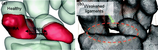
Fig. 5.5
The simulated SLD and SLAC were shown in (a) where the RA bones were in red and the transparent bones represent the healthy bones. The simulated effect of weakened and torn ligaments was also shown (b)
5.1.6 Simulation of Dislocation of the Scaphoid in the Palmar Direction
Dislocation of the scaphoid in the palmar direction was due to the radial insertion of the Testut ligament synovialitis has caused bone loss and the possibility of so-called Mannerfelt crypt [2]. This is also one of the criteria of the SLAC [5]. The simulation (Fig. 5.6) was performed by rotating radially the scaphoid (center of scaphoid as COR) and palmarly (the proximal end ulnar direction as COR) for 16.8 and 22.3°, respectively.
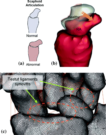
Fig. 5.6
Information from literature on the rotatory subluxation of the scaphoid, producing incongruent loading at the radioscaphoid facet [5] was used to perform the simulation (a).The simulated scaphoid dislocation in the palmar direction from sagittal view (b) and palmar view (c). The RA bones were in red and the transparent bones represent the healthy bones
5.1.7 Simulation of Hand Scoliosis
Hand scoliosis occurs due to tendon rupture. This mechanism ends in a changed axis of the wrist to the ulna with a consecutive rotation of the metacarpal bones in the radial direction [2]. Hand scoliosis was simulated by dislocating 7.23 mm all carpus excluding the scaphoid towards ulnar and rotating radially 10° of all metacarpals with the center of the radius as the COR. This mechanism resulted in a changed axis of the wrist to the ulnar [2] (Fig. 5.7).
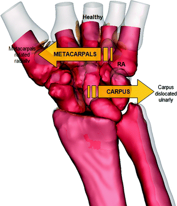
Fig. 5.7
The dislocation of carpus towards ulnar and rotation of metacarpals radially due to tendon rupture resulted in hand scoliosis. The RA bones were in red while transparent bones depicting the normal healthy wrist
5.1.8 Simulation of Reduction of Contact Between the Lunate and the Radius
As revealed by Trieb et al., the contact between the lunate and the radius was decreased in rheumatic wrist [2]. It was due to the dislocation of the proximal row or the carpal bones towards ulnar. This condition was simulated through translation of 7 mm of the lunate towards ulnar direction (reference was positioned at the center of the distal ulna) resulting in decreasing of the contact between the radius and the lunate (Fig. 5.8).
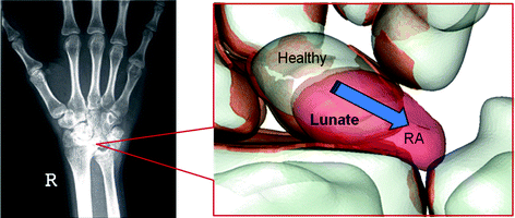
Fig. 5.8
The palmar view of the carpus where the RA bones were in red while transparent bones depict the normal healthy wrist. The figure shows the translation of the lunate towards ulnar direction resulting in the decreasing of the contact between the radius and the lunate
5.1.9 Simulation of Bone Erosion
Bone erosion was simulated by using Boolean operation (subtraction) after assuring accuracy of the bone’s position (Fig. 5.9). Sharp edges due to eroded bone were manually simulated by utilising local smoothing algorithm tool. Bone erosion was regularly occurred in the rheumatic wrist attributed to the inflammation of the synovial fluid and deterioration of the joint constraint [1–3, 6–8]. The differences of volumes between the healthy and the RA bones were summarised in Table 5.2.
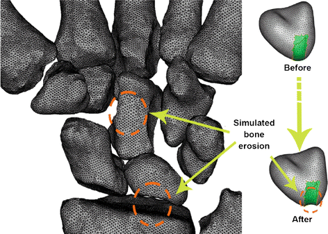
Fig. 5.9
The effect of bone erosion was shown in this exploded view of the RA model (left)
Table 5.2
Information on the difference between the healthy and RA wrist after simulated bone erosion
No. | Bone | Healthy (mm3) | RA (mm3) | Bone Erosion (%) |
|---|---|---|---|---|
1 | 1MC | 3852.1 | 3766.0 | 2.2 |
2 | 2MC | 2423.8 | 2312.0 | 4.6 |
3 | 3MC | 2946.1 | 2600.1 | 11.7 |
4 | 4MC | 1678.9 | 1603.6 | 4.5 |
5 | 5MC | 2414.8 | 2223.2 | 7.9 |
6 | Trapezium | 1646.6 | 1336.9 | 18.8 |
7 | Trapezoid | 1177.1 | 926.5 | 21.3 |
8
Related posts:Stay updated, free articles. Join our Telegram channel
Full access? Get Clinical Tree
 Get Clinical Tree app for offline access

|


