Hip dysplasia usually has associated femoral bone deformity. When total hip arthroplasty is considered, femoral bone deformity mandates special considerations in femoral reconstruction. The common deformities in the dysplastic femur can pose technical challenges leading to difficult exposure, increased risk of fracture, implant malposition, hip instability/impingement, and compromise of implant fixation. Management of leg length on the femoral side is also a frequent challenge. Modern surgical techniques and implants have provided solutions to address the complex issues that arise in femoral reconstruction of the dysplastic hip, allowing successful and durable outcomes in total hip arthroplasty.
- •
The common deformities in the dysplastic femur include hypoplasia, excessive neck anteversion, a valgus neck-shaft angle, metaphyseal-diaphyseal mismatch, and a posteriorly displaced greater trochanter.
- •
Depending on the degree of hip dysplasia, where the cup is placed, and length of the opposite leg, management of leg length on the femoral side is also a frequent challenge.
- •
Although often thought of as an acetabular deformity, hip dysplasia also usually has associated femoral bone deformity.
Introduction
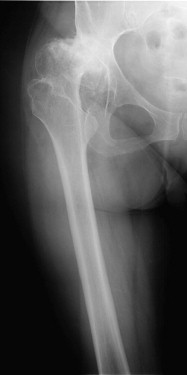
Developmental dysplasia of the hip (DDH) is one of the most common conditions leading to secondary arthritis of the hip ( Fig. 1 ). After nonarthroplasty options are exhausted, many patients with symptomatic arthritis secondary to dysplasia become candidates for total hip replacement. There is a predictable spectrum of femoral and acetabular abnormalities that must be considered when contemplating total hip arthroplasty (THA) in the patient with DDH. This article presents the specific considerations related to femoral reconstruction during THA in patients with hip dysplasia.
Milder cases of bone deformity present little difference from standard approaches to femoral reconstruction. In contrast, more severe bone and soft tissue deformities can present a multitude of issues including hypoplasia of the femur, excessive femoral anteversion, a valgus neck-shaft angle, metaphyseal-diaphyseal mismatch, a posteriorly displaced greater trochanter, and challenges related to leg length.
Preoperative radiographs with magnification markers should be obtained on all dysplastic patients who are being considered for THA. The surgeon should look carefully at these films to assess for signs associated with the dysplastic femur. Specific elements to scrutinize are the amount of proximal migration of the femoral head, the location of the greater trochanter, the size of the femoral canal, and overall femoral morphology. It is difficult to assess the amount of femoral anteversion without the use of three-dimensional imaging, although marked abnormalities frequently are identifiable on plain films. Although it is not the routine practice of the authors, three-dimensional imaging may be obtained if understanding the amount of anteversion before surgery will assist the surgeon.
DDH may be classified according to several systems. The most common is that of Crowe and colleagues based on the extent of radiographic proximal migration of the femoral head. The extent of subluxation does correlate with the degree of femoral deformity, but not predictably. Robertson and colleagues were the first to use computed tomography (CT) scans to study the morphology of the dysplastic femur. They evaluated 24 hips with DDH and found that femoral anteversion extended down through the metaphysis to the level of the lesser trochanter. In accordance with previous studies, in the diaphysis, the femoral canal was wider in the anterior to posterior direction than in the medial to lateral direction. The extent of femoral deformity was not significantly influenced by the extent of disease as classified by Crowe and colleagues. Sugano and colleagues also used three-dimensional CT reconstructions to study the morphology of 35 dysplastic hips compared with a matched cohort. Compared with the control group, the dysplastic femurs had shorter necks, smaller intramedullary canals, and 10° to 14° more anteversion independent of the amount of subluxation of the hip. Noble and colleagues reported on 154 women with DDH and found that patients with more advanced subluxation (Crowe III and IV) had increased hypoplasia of the femur, deformity of the femoral head, and straighter femoral canals with thinner cortices. However, even those femurs with mild subluxation (Crowe I) had measurable alterations in the anatomy of their proximal femur. Argenson and colleagues reviewed the radiographs and CT scans of 83 hips with DDH and compared femoral morphology with a control group. The intramedullary femoral canal had reduced dimensions in the mediolateral and anteroposterior planes in all patients with DDH compared with the control group. There was no correlation between femoral morphology and Crowe class.
Technical challenges unique to femoral reconstruction in DDH
The anatomic abnormalities of the dysplastic femur are numerous and present reconstructive challenges to the surgeon. In addition, patients frequently have undergone previous nonarthroplasty procedures that have altered their already distorted anatomy. It is important to be familiar with the patient’s previous procedures, because sometimes canal realignment procedures must take place concomitantly with THA ( Fig. 2 ). For example, valgus subtrochanteric (Schantz) osteotomy, can present considerable difficulty in femoral reconstruction during THA.
Exposure
Several operative approaches can be performed safely for THA in patients with hip dysplasia. For mild dysplasia, adequate exposure usually can be obtained through any of the popular approaches to the hip including direct anterior, anterolateral, or the posterior approach, based on surgeon preference. Subtrochanteric osteotomy may be used to adjust femoral anteversion or to shorten the femur. This osteotomy also provides exposure of the acetabulum in cases of high hip dislocation. Greater trochanteric osteotomy or a trochanteric slide may be used in selected situations in which the hip cannot be dislocated or exposed with a traditional soft tissue exposure or in which greater trochanteric repositioning is desirable to restore hip biomechanics.
Restoration of abductor function is crucial to the success of THA in the dysplastic hip. The proximal displacement of the femoral head relative to the true acetabulum and excessive femoral neck anteversion can cause severe distortion of the length and direction of the abductor muscle fibers. This distortion must be considered during the operative approach to avoid proceeding through improper planes of dissection. The abductors are often underdeveloped and more transverse in nature than normal. This distortion of the abductor muscles can severely affect their function, making them less efficient. Restoration of the hip to its true hip center and correction of femoral neck anteversion can often provide more normal abductor anatomy and function. Specific techniques to restore length and position of the greater trochanter are discussed later.
Canal Diameter and Shape
Charnley and Feagin showed that the canal of the dysplastic femur is often extremely narrow and is frequently an exaggerated oval in cross section, wider anterior to posterior than medial to lateral. This shape complicates canal preparation. It is imperative to measure the size of the intramedullary canal before surgery to ensure that appropriately sized and shaped femoral components are available at the time of surgery. Proper component sizing minimizes the risk of fracture (or perforation) during canal preparation and seating of the femoral component.
Anteversion
Femoral neck anteversion is defined as the angle between the transverse axis of the knee and the transverse axis of the femoral neck. Femoral anteversion varies widely in patients with DDH. Three-dimensional imaging is a reliable method to accurately assess the amount of femoral neck anteversion, which can reach as much as 90°. Surgeons must be prepared to deal with a varying degree of anteversion encountered at the time of surgery. If excessive femoral anteversion in the dysplastic femur is not recognized, excessive anteversion of the femoral component may ensue and can lead to intraprosthetic impingement, (anterior) hip instability, or an internal rotation gait that is not well tolerated by patients.
Minor or moderate degrees of femoral anteversion abnormality can be corrected during THA using certain standard implants and methods of fixation. In cases of more severe anteversion abnormalities, there are 2 effective methods of correction: subtrochanteric osteotomy; and special, uncemented stems (either modular or conical with flutes). With a subtrochanteric osteotomy, the goal is to rotate the proximal fragment such that the metaphyseal flare and the greater trochanter are placed into a more anatomic position. This position helps restore anatomic and functional characteristics of the abductors and also minimizes the risk of impingement of the trochanter on the pelvis. Furthermore, this repositions the metaphyseal flare to a position that accommodates the triangular metaphyseal portion of a femoral component.
Leg Length
In many patients with DDH, it is desirable and/or necessary to lengthen the operative extremity by placing the acetabular component in an anatomic location, particularly in unilateral cases with a short leg or bilateral cases with higher levels of subluxation/dislocation. One concern associated with lengthening that has been shown repeatedly is risk of palsies of the sciatic or femoral nerves.
Dunn and Hess suggested that it was safe to lengthen the limb by 5 to 7 cm in the setting of dysplasia. In their series, 13 of 22 hips were lengthened by at least 5 cm (range 5–9.2 cm). None of the patients in this series with limbs lengthened by more than 5 cm experienced a nerve palsy. The 1 patient who developed a (sciatic) nerve palsy was lengthened by 4 cm.
In a report by Edwards and colleagues, 23 hips out of 614 consecutive THAs experienced either a peroneal or sciatic nerve palsy. Twenty-two percent of these patients had an underlying diagnosis of DDH. This study was the first to show a relationship between the amount of lengthening and the development of a nerve palsy. Peroneal nerve palsies were associated with limb lengthening of 3.8 cm or less, and sciatic nerve palsies were associated with lengthening of 4.0 cm or more. These data have yet to be corroborated.
In their review of 3126 consecutive THAs, Schmalzried and colleagues found a 5.2% incidence of nerve palsies in patients with DDH. Of the 9 patients with DDH who experienced a nerve palsy, 6 had their limbs lengthened by more than 3.0 cm. Of all 52 patients who experienced a nerve palsy in this study, 13 patients had their operative extremity lengthened by more than 2.5 cm. The investigators concluded that limb lengthening is a major contributor to nerve palsy after THA, but failed to identify the specific parameters of lengthening that should be avoided.
Farrell and colleagues reported on 27,004 consecutive primary THAs performed at Mayo Clinic (not all for DDH) and found that, in 43 patients with a (sciatic, peroneal, or femoral) nerve injury, overall length of the extremity was increased by an average of 1.7 cm. A matched cohort without nerve injury had the operative limb lengthened by only 1.1 cm. A conditional, logistical regression analysis showed that patients with increased limb lengthening were at increased risk for nerve injury.
Although the amount of lengthening that causes nerve dysfunction is not known, if templating suggests that reducing the femur into an anatomically placed acetabular component lengthens the limb by more than 3 to 4 cm, the authors typically consider shortening of the femur to reduce the amount of leg lengthening. Intraoperative electromyographic monitoring may also be considered in these cases, although its value in reducing risk of nerve palsy is unproven.
Related posts:
 Strategies to Improve Nonoperative Childhood Management
Surgical Treatment of Hip Dysplasia in Children and Adolescents
Bearing Surface Considerations for Total Hip Arthroplasty in Young Patients
Technical Considerations in Total Hip Arthroplasty After Femoral and Periacetabular Osteotomies
Surgical Treatment of Hip Dysplasia in Children and Adolescents
Bearing Surface Considerations for Total Hip Arthroplasty in Young Patients
Strategies to Improve Nonoperative Childhood Management
Surgical Treatment of Hip Dysplasia in Children and Adolescents
Bearing Surface Considerations for Total Hip Arthroplasty in Young Patients
Technical Considerations in Total Hip Arthroplasty After Femoral and Periacetabular Osteotomies
Surgical Treatment of Hip Dysplasia in Children and Adolescents
Bearing Surface Considerations for Total Hip Arthroplasty in Young Patients
Stay updated, free articles. Join our Telegram channel

Full access? Get Clinical Tree


