Excision of Talocalcaneal Coalition
David Scher
DEFINITION
A talocalcaneal coalition is an abnormal connection between the talus and the calcaneus that limits subtalar motion.
As is the case for calcaneonavicular coalitions, which are described in the prior chapter, talocalcaneal coalitions typically result in a rigid flatfoot that is sometimes painful.
ANATOMY
Talocalcaneal coalitions occur within the subtalar joint, most commonly involving the middle facet.9
These connections can be bony, cartilaginous, or fibrous and can involve any amount of the joint.
The size of the coalition is described with respect to the percentage of the entire subtalar joint that is coalesced.9
PATHOGENESIS
Like calcaneonavicular coalitions, the cause of talocalcaneal coalitions remains unknown, but they may be the result of failure of segmentation during fetal development.1
Although they are presumed to be congenital in nature, symptoms typically do not appear until early adolescence, ages 12 to 16 years.7, 10
It is unclear why some coalitions become painful. One theory suggests the possibility of altered talar joint kinematics placing additional stress on adjacent joints. Another is the development of microfractures or stress fractures through the coalition over time, rendering them painful.1
NATURAL HISTORY
Most talocalcaneal coalitions are asymptomatic.6
They may result in the development of a rigid flatfoot, characterized by valgus alignment of the heel, abduction of the forefoot, loss of the arch, and failure of the arch to reconstitute on toe-rise or when non-weight bearing.
PATIENT HISTORY AND PHYSICAL FINDINGS
Patients typically describe pain in the foot that is activity related; it is exacerbated by walking on uneven surfaces and relieved by rest.
This pain may be generalized to the midfoot and hindfoot or can be specifically localized to the medial aspect of the hindfoot and ankle.
Patients may also complain of lateral pain at the tip of the fibula.
There may be a history of progressively worsening out-toeing or loss of the arch.
The clinician should observe the patient’s gait for an antalgic pattern and torsional alignment, with specific attention to foot position during stance.
The patient’s foot alignment is examined; the heel may be in valgus alignment with the forefoot abducted.
The rigidity of the flatfoot is observed. Flexible flatfoot has a restoration of the arch on toe-rise; rigid flatfoot has no arch restoration. A rigid flatfoot is a sign of decreased subtalar motion and may indicate a tarsal coalition.
The physician should test for subtalar motion. The test is not specific for talocalcaneal coalition but is indicative of some process within the subtalar joint.
The physician should palpate over the medial aspect of the hindfoot, just plantar to the medial malleolus, in the region of the sustentaculum tali. Tenderness in this region may be indicative of a middle facet talocalcaneal coalition.
IMAGING AND OTHER DIAGNOSTIC STUDIES
Plain radiographs should be obtained in an attempt to identify the coalition and assess foot alignment. These should include anteroposterior (AP), lateral, oblique, and Harris axial views (FIG 1A,B).
A talocalcaneal coalition is best seen on the Harris axial view, but it may be difficult to obtain the exact orientation to adequately visualize the middle facet.
On the lateral view, there may be a continuous C-shaped line along the talar dome and into the posterior facet (C-sign).8
On the AP and oblique view, one may identify other concurrent coalitions.
Standing AP and lateral radiographs and a Saltzman hindfoot alignment view can be useful for assessing foot alignment, especially hindfoot valgus.
A computed tomography (CT) or magnetic resonance imaging (MRI) scan is mandatory to clearly visualize the coalition and determine the percentage of the subtalar joint that is involved (FIG 1C).9
An MRI may be useful if the diagnosis is equivocal and a cartilaginous or fibrous coalition is suspected.
DIFFERENTIAL DIAGNOSIS
Flexible flatfoot
Subtalar arthritis
Other tarsal coalition (calcaneonavicular or less common ones)
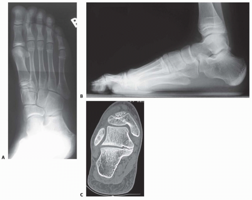
FIG 1 • A. The oblique radiograph confirms the absence of a concurrent calcaneonavicular tarsal coalition. B. On the lateral radiograph, a C-sign is visible as a confluent line around the posterior margin of the talar body and the posterior aspect of the calcaneus just beneath the posterior facet of the subtalar joint. This sign is often associated with a talocalcaneal tarsal coalition. C. CT scanning can be used to visualize the coalition and determine the percentage of the subtalar joint that is involved.
Tumor or infection involving the subtalar joint
Idiopathic rigid flatfoot
NONOPERATIVE MANAGEMENT
Nonoperative management is indicated for all patients with talocalcaneal coalition at first presentation.
Painless coalitions need no treatment.
The initial treatment for painful talocalcaneal coalitions is activity modification, anti-inflammatory medication, or immobilization in a short-leg walking cast.
SURGICAL MANAGEMENT
The indication for surgical management is persistence of pain despite nonoperative management.
The main goals of treatment are, primarily, elimination of pain and restoration of function.
Restoration of subtalar motion is a secondary goal.
Restoration of arch height is unlikely following excision of a talocalcaneal coalition.
Preoperative Planning
All imaging studies are reviewed.Related posts:

Stay updated, free articles. Join our Telegram channel

Full access? Get Clinical Tree


