Fig. 8.1
Bilateral coxa vara with neck shaft angle approaching 106°
It has been described in many different ways over the years, but we shall use the following classification [1]:
- 1)
Developmental (previously named Infantile or Cervical)
- 2)
Congenital (associated with congenital short femur)
- 3)
Acquired (encompassing traumatic and those associated with skeletal dysplasias)
For the purpose of this chapter, we shall concentrate on Developmental coxa vara, as treatment of the various other types of coxa vara will depend on the heterogeneous underlying causes of the deformity.
Developmental coxa vara in this sense occurs at the level of the physis, whereas the other causes occur distal to the physis.
Background
The finding of Developmental coxa vara (DCV) was first described by the Italian Fiorani in 1881 as a “bending” of the femoral neck [2]. He believed that the origin was rachitic. In 1881 Muller described a similar appearance in adolescents, but the terminology coxa vara was attributed to the condition described by Hofmeister in 1894 [3, 4].
Hoffa in 1905 examined pathological specimens of the proximal femur in affected individuals and concluded that the aetiology was unlikely to be either rickets or trauma and instead introduced the idea of a developmental condition [5].
In 1960 Pylkkanen performed a detailed literature search of the available information relating to coxa vara as well as adding weight to the pathophysiological knowledge of the influencing processes [6]. In Scandinavia, Johanning reported an incidence of 1 in 25,000 live births in the early 1950s [6]. Males and females are equally affected by this condition and there appears to be no difference in the frequency of the side involved. It may be bilateral in up to one third of individuals.
Normal Femoral Development
The proximal femur in the neonate is completely cartilaginous, with the recognisable shape of the femoral head and the trochanters determined by the enchondral ossification at the junctions between the metaphysis and the epiphyseal plates. The physeal plate gradually migrates proximally and divides into 2 separate plates, one for the femoral head and the second one for the greater trochanter. The medial portion of the cervical physis progresses early, resulting in elongation of the femoral neck. An ossification centre appears within the femoral head somewhere between 4 and 7 months after birth and can then be seen on x-ray. The more lateral greater trochanteric apophysis starts to ossify at about 4 years of age. The proximal femoral growth plate accounts for one third of the longitudinal growth of the femur.
Relative growth of these physes results in the changing morphology of the proximal femur. At birth, the normal neck-shaft angle is about 150° and in adulthood it is approximately 127° (often decreasing to 120° by the 7th decade). In a similar fashion, anteversion of the femoral neck may be as much as 40° at birth. By adulthood the normal femoral anteversion ranges from 4° to 25°.
Pathophysiology
Over the past century, many different theories have been suggested as causes of coxa vara, including Staphylococcal Albus infections, bending of the neck secondary to pressure within the uterus, rickets and AVN-like changes. In 1928 Fairbank described a triangular piece of bone in the distal portion of the medial femoral neck as a potential cause of the problem [7].
Following work by Zimmerman (1938) and others, it has become clear that the condition does not exist at birth, but develops after the perinatal period as a result of a defect in the enchondral ossification of the medial femoral neck [8]. Hence the nomenclature of “developmental” coxa vara.
On histopathological examination of specimens obtained in Helsinki and compared to normal individuals, there were abnormalities of varying severity found in 3 main areas [6]:
- a)
Changes in ossification at the physeal cartilaginous plate, with disorganisation and irregularity of columns of cells
- b)
Changes in the adjacent metaphysis, including thin, irregular trabeculae and lack of proper ossification
- c)
Fibrous connective tissue infiltrating the bone marrow of the metaphysis and within the cartilaginous tissue
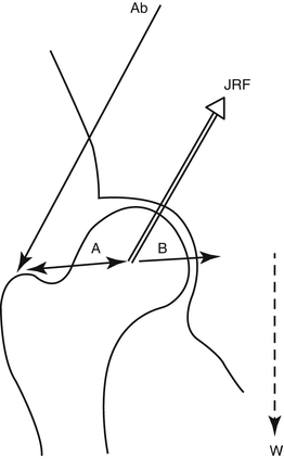
Deformation of the femoral neck can occur because of reduced strength in the region of the medial femoral neck because of bony and cartilaginous abnormalities. Once the neck-shaft angle reduces, the greater trochanter migrates towards the ilium and abduction is reduced. The higher level of the greater trochanter increases the abductor lever arm, reducing the joint reaction force crossing the hip in single leg stance. The joint reaction force required to maintain a horizontal pelvis during single stance gait decreases (Fig. 8.2).


Fig. 8.2
Free body diagram of the hip in coxa vara. Joint reaction force is the force generated within a normal hip joint. This is a balance between the body weight moment arm (B) and the body weight (W), and the abductor moment arm (A) and abductor tension (Ab)
In the normal hip, the force transmitted to the proximal femoral neck would include a net tension force at the superior or lateral cortex and a net compressive force at the inferior or medial cortex.
However, the shearing and bending forces on the neck will increase with increasing varus deformity as the physis changes its position from horizontal to more vertical, with restriction of growth on the medial physis relative to the lateral physis. The more vertical position of the proximal femoral physis would increase not only the sheer component of the hip force but also the net medial compressive force on the metaphyseal bone of the femoral neck. These forces overwhelm the mechanical strength of the abnormally ossified bone in this area. This may lead to a relentless and progressive cycle of deformity.
Stay updated, free articles. Join our Telegram channel

Full access? Get Clinical Tree


