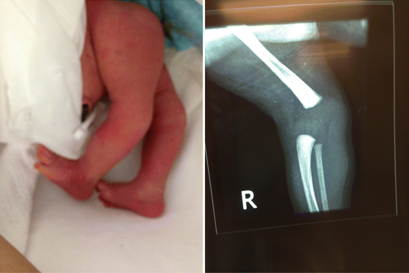
Fig. 11.1
Syndromic knees and hips dislocations
The pathology of CDK is similar in most cases with short quadriceps tendon, tight anterior knee joint capsule and hypoplastic suprapatellar pouch with anteriorly subluxed hamstrings. However, the severity of these changes is exaggerated in complex cases and associated with secondary changes in bones and ligaments in long standing deformities [4].
The clinical presentation of knee recurvatum leads easily to early diagnosis and can be confirmed by radiography [5, 6]. The relation between the tibia and femur clinically and radiographically allows for a simple classification of CDK into recurvatum, subluxation and dislocation (Leveuf’s Classification). The simple form of genu recurvatum is usually related to fetal molding due to oligohydramnios or breech position as been suggested by Haga et al. [7].
The other classification system was proposed by Finder in 1964 dividing CDK into five types according to the severity of the dislocation and its complexity. Type 1 being physiologic hyperextension up to 20° and type 5 being a complex variant including mixed categories of congenital diseases such as Ehlers-Danlos syndrome and Arthrogryposis [8] (Table 11.1, Fig. 11.2).

Table 11.1
Finder classification
Type I | Physiologic hyperextension up to 20° considered within normal limits. Usually disappears by age 8 years (Fig. 11.2) |
Type II | Simple hyperextension, a continuation of type I into adult life |
Type III | Anterior subluxation with knee hyperextension up to 90° and resisted flexion beyond neutral |
Type IV | Dislocation of the knee with the proximal tibia migrated upward and anteriorly |
Type V | Complex variants including a mixed category of congenital diseases such as Ehlers-Danlos syndrome and arthrogryposis |

Fig. 11.2
Congenital knee dislocation, Finder type I
How and When Should We Treat Congenital Dislocation of the Knee?
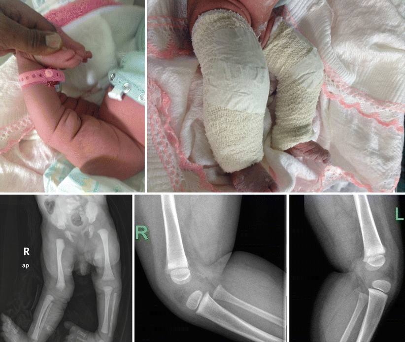
CDK is a rare entity and the existing studies in the literature are case series and case reports (Level IV and V). Most of the studies initiate management with non-surgical treatment followed by surgical intervention for failed non-surgical cases. Many modalities can be used for non-surgical treatment including the use of serial casting (Fig. 11.3), pavlik harness, skin and skeletal traction or observation. Surgical interventions include minimally invasive quadriceps tenotomy, open quadricepsplasty with joint capsule release and femoral shortening.


Fig. 11.3
Congenital knee dislocation treated with serial casting
Early Versus Late Reduction
Different authors have reported variable success rates following non-surgical treatment of CDK. However, most authors agree that early treatment has an impact on the outcome in non-syndromic cases of CDK [9, 10]. Haga et al. suggested waiting 1 month for spontaneous reduction of CDK not associated with clubfoot, AMC or Larsen’s syndrome [7].
Johnson et al. reviewed 17 patients with a follow-up of 11 years. Nine patients were treated non-surgically with good and fair results in 7 patients; all but 2 were less than 2 years of age when treatment was started with unilateral involvement. The role of arthrography was investigated in this study and found to be questionable as a technique to predict the response to close reduction because it was done usually before open reduction. Furthermore, a constant finding was ablation of the suprapatellar pouch in all surgical cases [11].
Meyer reviewed 68 patients and found that treatment was successful in 81 % of patients if performed before the age of 3 months. The rate of success dropped to 33 % if treatment was initiated between the ages of 3 and 6 months. [12].
Nogi and MacEwen successfully treated all but three of seventeen patients (excluding those with arthrogryposis or neuromuscular disorders) with manipulation and serial casting or Pavlik harness immediately after birth. The three failures occurred in two of delayed treatment cases and one with pseudo-reduction of the knee joint [13].
Ferris and Aichroth treated 19 CDK cases, nine of them treated non-surgically. Excellent and good results were achieved in five of the nine patients when the treatment started before 3 months of age and poor outcomes in syndromic patients with late treatment [6].
Isolated Versus Syndromic CDK
Ooishi et al. reviewed the results of 19 patients treated between 1972 and 1990. Twelve patients with isolated CDK and the remaining were syndromic patients (one with Larsen’s syndrome and the other six with AMC). Non-surgical treatment started after birth in all patients. The isolated CDK cases reduced and all of the knees except one showed normal knee joint development. In contrast, the same treatment resulted in limited effect and residual subluxation in all the AMC and Larsen’s syndrome patients. Three of the AMC patients underwent open reduction with quadricepsplasty [4]. Although treatment started early, non-surgical treatment failed in the syndromic and complicated cases in this study. Roy and Crawford described a percutaneous recession of the quadriceps mechanism through three stab incisions for Finder type 5 cases that are syndromic and associated with multiple anomalies. The advantage of such an approach is the avoidance of scar tissue and other complications potentially caused with more extensive surgery. The mean age of the patients at the time of surgery was 18 days, which further emphasizes the importance of early intervention for complex cases [14].
Percutaneous Tenotomy Versus Open Quadricepsplasty
Despite the general consensus on the necessity of surgical correction of CDK for unsuccessful cases to non-surgical treatment, there is no consensus regarding the type of surgical intervention. The fundamental pathological feature in CDK involves the quadriceps tendon and anterior joint capsule. Hence, most of the surgical procedures address these pathological changes to facilitate a joint reduction of the knee.
Curtis and Fisher described a long anterolateral approach with extensive mobilization of the quadriceps muscle, tendon lengthening via Z-plasty or an inverted V-incision, and an anterior capsulotomy of the knee [5]. Although successful in reducing the dislocated knee, this extensive approach unsurprisingly causes extensive scarring of the extensor mechanism; adhesions and wound complications. These complications often result in unsatisfactory outcomes, therefore this procedure was recommended for recurrence or severe cases with failed previous treatment [15].
Stay updated, free articles. Join our Telegram channel

Full access? Get Clinical Tree


